SEARCH
検索詳細

伊藤 俊樹バイオシグナル総合研究センター教授
研究者基本情報
■ 学位■ 研究ニュース
■ 研究分野
■ 委員歴
研究活動情報
■ 受賞■ 論文
- Water freeze-drying has traditionally been considered an inappropriate drying method for biological samples for scanning electron microscopy (SEM) because ice crystals can potentially form and damage cell structures. Chemical fixation is also considered essential for the SEM observation of soft cells, such as protozoa and animal cells. We succeeded in preparing specimens of freshwater protists for SEM using simple water freeze-drying without chemical fixation. Specifically, a suspension of live cells in water was frozen by contact with a - 80 °C copper block using an outer cylinder that ensured precise planar contact. This significantly reduced artifact formation and increased the success rate from ~ 10% to 20-30%. Transmission electron microscopy (TEM) observations confirmed that intracellular ice crystal formation was significantly suppressed in approximately 5% of Paramecium cells, representing a notable improvement over conventional methods. This method also shortens preparation time to 2-3 h and enables high-resolution SEM imaging of various organisms. Overall, based on these findings, we standardized a simple and low-artifact method using water freeze-drying as a novel and improved technique for SEM sample preparation.2025年12月, Scientific reports, 英語, 国際誌研究論文(学術雑誌)
- 2025年12月, Life Science Alliance研究論文(学術雑誌)
- Elsevier BV, 2025年06月, Biochemical and Biophysical Research Communications, 766, 151846 - 151846研究論文(学術雑誌)
- Osteoclasts are multinucleated giant cells that are formed by the fusion of precursor cells. Cell–cell fusion is mediated by membrane protrusion driven by actin reorganization, but the role of membrane mechanics in this process is unknown. Utilizing live-cell imaging, optical tweezers, manipulation of membrane-to-cortex attachment (MCA), and genetic interference, we show that a decrease in plasma membrane (PM) tension is a mechanical prerequisite for osteoclast fusion. Upon RANKL-induced differentiation, ezrin expression in fusion progenitor cells is reduced, resulting in a decrease in MCA-dependent PM tension. A forced elevation of PM tension by reinforcing the MCA conversely suppresses cell–cell fusion. Mechanistically, reduced PM tension leads to membrane protrusive invadosome formation driven by membrane curvature-inducing/sensing BAR proteins, thereby promoting cell–cell fusion. These findings provide insights into the mechanism of cell–cell fusion under the control of membrane mechanics.Rockefeller University Press, 2025年05月, Journal of Cell Biology, 224(7) (7)研究論文(学術雑誌)
- Elsevier BV, 2023年04月, Current Opinion in Cell Biology, 81, 102173 - 102173[査読有り][招待有り]研究論文(学術雑誌)
- Abstract Epithelial cells provide cell-cell adhesion that is essential to maintain the integrity of multicellular organisms. Epithelial cell-characterizing proteins, such as epithelial junctional proteins and transcription factors are well defined. However, the role of lipids in epithelial characterization remains poorly understood. Here we show that the phospholipid phosphatidylinositol (4,5)-bisphosphate [PI(4,5)P2] is enriched in the plasma membrane (PM) of epithelial cells. Epithelial cells lose their characteristics upon depletion of PM PI(4,5)P2, and synthesis of PI(4,5)P2 in the PM results in the development of epithelial-like morphology in osteosarcoma cells. PM localization of PARD3 is impaired by depletion of PM PI(4,5)P2 in epithelial cells, whereas expression of the PM-targeting exocyst-docking region of PARD3 induces osteosarcoma cells to show epithelial-like morphological changes, suggesting that PI(4,5)P2 regulates epithelial characteristics by recruiting PARD3 to the PM. These results indicate that a high level of PM PI(4,5)P2 plays a crucial role in the maintenance of epithelial characteristics.Springer Science and Business Media LLC, 2022年12月, Nature Communications, 13(1) (1)[査読有り]研究論文(学術雑誌)
- Abstract Angiogenesis is regulated in coordinated fashion by chemical and mechanical cues acting on endothelial cells (ECs). However, the mechanobiological mechanisms of angiogenesis remain unknown. Herein, we demonstrate a crucial role of blood flow-driven intraluminal pressure (IP) in regulating wound angiogenesis. During wound angiogenesis, blood flow-driven IP loading inhibits elongation of injured blood vessels located at sites upstream from blood flow, while downstream injured vessels actively elongate. In downstream injured vessels, F-BAR proteins, TOCA1 and CIP4, localize at leading edge of ECs to promote N-WASP-dependent Arp2/3 complex-mediated actin polymerization and front-rear polarization for vessel elongation. In contrast, IP loading expands upstream injured vessels and stretches ECs, preventing leading edge localization of TOCA1 and CIP4 to inhibit directed EC migration and vessel elongation. These data indicate that the TOCA family of F-BAR proteins are key actin regulatory proteins required for directed EC migration and sense mechanical cell stretching to regulate wound angiogenesis.Springer Science and Business Media LLC, 2022年12月, Nature Communications, 13(1) (1)[査読有り]研究論文(学術雑誌)
- Abstract Malignancy is associated with changes in cell mechanics that contribute to extensive cell deformation required for metastatic dissemination. We hypothesized that the cell-intrinsic physical factors that maintain epithelial cell mechanics could function as tumor suppressors. Here we show, using optical tweezers, genetic interference, mechanical perturbations, and in vivo studies, that epithelial cells maintain higher plasma membrane (PM) tension than their metastatic counterparts and that high PM tension potently inhibits cancer cell migration and invasion by counteracting membrane curvature sensing/generating BAR family proteins. This tensional homeostasis is achieved by membrane-to-cortex attachment (MCA) regulated by ERM proteins, whose disruption spontaneously transforms epithelial cells into a mesenchymal migratory phenotype powered by BAR proteins. Consistently, the forced expression of epithelial–mesenchymal transition (EMT)-inducing transcription factors results in decreased PM tension. In metastatic cells, increasing PM tension by manipulating MCA is sufficient to suppress both mesenchymal and amoeboid 3D migration, tumor invasion, and metastasis by compromising membrane-mediated mechanosignaling by BAR proteins, thereby uncovering a previously undescribed mechanical tumor suppressor mechanism.Springer Science and Business Media LLC, 2021年10月, Nature Communications, 12(1) (1)[査読有り]研究論文(学術雑誌)
- At the initial stage of carcinogenesis, cell competition often occurs between newly emerging transformed cells and the neighboring normal cells, leading to the elimination of transformed cells from the epithelial layer. For instance, when RasV12-transformed cells are surrounded by normal cells, RasV12 cells are apically extruded from the epithelium. However, the underlying mechanisms of this tumor-suppressive process still remain enigmatic. We first show by electron microscopic analysis that characteristic finger-like membrane protrusions are projected from both normal and RasV12 cells at their interface. In addition, FBP17, a member of the F-BAR proteins, accumulates in RasV12 cells, as well as surrounding normal cells, which plays a positive role in the formation of finger-like protrusions and apical elimination of RasV12 cells. Furthermore, cdc42 acts upstream of these processes. These results suggest that the cdc42/FBP17 pathway is a crucial trigger of cell competition, inducing "protrusion to protrusion response" between normal and RasV12-transformed cells.2021年09月, iScience, 24(9) (9), 102994 - 102994, 英語, 国際誌[査読有り]研究論文(学術雑誌)
- We characterized the size, distribution, and fluidity of microdomains in a lipid bilayer containing phosphatidylinositol (PI) and revealed their roles during the two-dimensional assembly of a membrane deformation protein (FBP17). The morphology of the supported lipid bilayer (SLB) consisting of PI and phosphatidylcholine (PC) on a mica substrate was observed with atomic force microscope (AFM). Single particle tracking (SPT) was performed for the PI+PC-SLB on the mica substrate by using the diagonal illumination setup. The AFM topography showed that PI-derived submicron domains existed in the PI+PC-SLB. The spatiotemporal dependence of the lateral lipid diffusion obtained by SPT showed that the microdomain had lower fluidity than the surrounding region and worked as the obstacles for the lipid diffusion. We observed the two-dimensional assembly of FBP17, which is one of F-BAR family proteins included in endocytosis processes and has the function generating lipid bilayer tubules in vitro. At the initial stage of the FBP17 assembly, the PI-derived microdomain worked as a scaffold for the FBP17 adsorption, and the fluid surrounding region supplied FBP17 to grow the FBP17 domain via the lateral molecular diffusion. This study demonstrated an example clearly revealing the roles of two lipid microregions during the protein reaction on a lipid bilayer.2021年05月, Membranes, 11(5) (5), 339, 英語, 国際誌[査読有り]研究論文(学術雑誌)
- Wiley, 2021年05月, FEBS Letters, 595(9) (9), 1303 - 1312[査読有り]研究論文(学術雑誌)
- Elsevier BV, 2021年03月, Biochemical and Biophysical Research Communications, 543, 15 - 22[査読有り]研究論文(学術雑誌)
- Ubiquitinated membrane proteins such as epidermal growth factor receptor (EGFR) are delivered to early endosomes and then sorted to lysosomes via multivesicular bodies (MVBs) for degradation. The regulatory mechanism underlying formation of intralumenal vesicles en route to generation of MVBs is not fully understood. In this study, we found that SH3YL1, a phosphoinositide-binding protein, had a vesicular localization pattern overlapping with internalized EGF in endosomes in the degradative pathway. Deficiency of SH3YL1 prevents EGF trafficking from early to late endosomes and inhibits degradation of EGFR. Moreover, we show that SH3YL1 mediates EGFR sorting into MVBs in a manner dependent on its carboxy-terminal SH3 domain, which is necessary for the interaction with an ESCRT-I component, Vps37B. Taken together, our observations reveal an indispensable role of SH3YL1 in MVB-sorting and EGFR degradation mediated by ESCRT complexes.The Company of Biologists, 2019年10月, Journal of Cell Science[査読有り]研究論文(学術雑誌)
- Abstract Tension in cell membranes is closely related to various cellular events, including cell movement and morphogenesis. Therefore, modulation of membrane tension can be a new approach for manipulating cellular events. Here, we show that an amphipathic peptide derived from the influenza M2 protein (M2[45–62]) yields lamellipodia at multiple sites in the cell. Effect of M2[45–62] on cell membrane tension was evaluated by optical tweezer. The membrane tension sensor protein FBP17 was involved in M2[45–62]-driven lamellipodium formation. Lysine-to-arginine substitution in M2[45–62] further enhanced its activity of lamellipodium formation. M2[45–62] had an ability to reduce cell motility, evaluated by scratch wound migration and transwell migration assays. An increase in neurite outgrowth was also observed after treatment with M2[45–62]. The above results suggest the potential of M2[45–62] to modulate cell movement and morphology by modulating cell membrane tension.Springer Science and Business Media LLC, 2019年06月, Communications Biology, 2(1) (1)[査読有り]研究論文(学術雑誌)
- Elsevier BV, 2018年01月, Biochemical and Biophysical Research Communications, 495(1) (1), 1522 - 1527[査読有り]研究論文(学術雑誌)
- 2015年10月, PLOS ONE, 10(10) (10), e0141569 - e0141569[査読有り]研究論文(学術雑誌)
- 2015年06月, Nature Cell Biology, 17(6) (6), 749 - 758[査読有り]研究論文(学術雑誌)
- 2015年06月, Biochimica et Biophysica Acta (BBA) - Molecular and Cell Biology of Lipids, 1851(6) (6), 824 - 831[査読有り]研究論文(学術雑誌)
- 2014年06月, Cancer Research, 74(11) (11), 3054 - 3066[査読有り]研究論文(学術雑誌)
- 2014年03月, Genes to Cells, 19(3) (3), 177 - 197[査読有り]研究論文(学術雑誌)
- American Society for Cell Biology (ASCB), 2013年11月, Molecular Biology of the Cell, 24(21) (21), 3393 - 3405[査読有り]研究論文(学術雑誌)
- 2013年01月, Langmuir, 29(1) (1), 328 - 336[査読有り]研究論文(学術雑誌)
- 2013年01月, Journal of Cell Science[査読有り]研究論文(学術雑誌)
- 2013年01月, Journal of Biochemistry, 153(1) (1), 21 - 29[査読有り]研究論文(学術雑誌)
- 2012年12月, Molecular Biology of the Cell, 23(24) (24), 4689 - 4700[査読有り]研究論文(学術雑誌)
- 2012年07月, Molecular Biology of the Cell, 23(13) (13), 2593 - 2604[査読有り]研究論文(学術雑誌)
- 2012年07月, Molecular Biology of the Cell, 23(13) (13), 2481 - 2489[査読有り]研究論文(学術雑誌)
- 2012年04月, Journal of Lipid Research, 53(4) (4), 810 - 819[査読有り]研究論文(学術雑誌)
- 2012年01月, Seikagaku. The Journal of Japanese Biochemical Society, 84(1) (1), 5 - 17, 日本語, 国内誌[Mechanisms for the regulation of the plasma membrane curvature in endocytosis].[査読有り][招待有り]研究論文(学術雑誌)
- 2011年07月, Journal of Biochemistry, 150(1) (1), 83 - 93[査読有り]研究論文(学術雑誌)
- Reversible interactions between cytosolic proteins and membrane lipids such as phosphoinositides play important roles in membrane morphogenesis driven by actin polymerization. In this paper, we identify a novel lipid-binding module, which we call the SYLF domain (after the SH3YL1, Ysc84p/Lsb4p, Lsb3p, and plant FYVE proteins that contain it), that is highly conserved from bacteria to mammals. SH3YL1 (SH3 domain containing Ysc84-like 1) strongly bound to phosphatidylinositol 3,4,5-triphosphate (PI(3,4,5)P3) and several D5-phosphorylated phosphoinositides through its SYLF domain and was localized to circular dorsal ruffles induced by platelet-derived growth factor stimulation. Interestingly, SHIP2 (the PI(3,4,5)P3 5-phosphatase, src-homology 2–containing inositol-5-phosphatase 2) was identified as a binding partner of SH3YL1, and knockdown of these proteins significantly suppressed dorsal ruffle formation. Phosphatidylinositol 3,4-bisphosphate (PI(3,4)P2), which is mainly synthesized from PI(3,4,5)P3 by the action of SHIP2, was enriched in dorsal ruffles, and PI(3,4)P2 synthesis strongly correlated with formation of the circular membrane structure. These results provide new insight into the molecular mechanism of dorsal ruffle formation and its regulation by phosphoinositide metabolism.Rockefeller University Press, 2011年05月, Journal of Cell Biology, 193(5) (5), 901 - 916[査読有り]研究論文(学術雑誌)
- 2010年02月, Journal of Biological Chemistry, 285(9) (9), 6781 - 6789[査読有り]研究論文(学術雑誌)
- 2010年01月, Journal of Human Genetics, 55(1) (1), 42 - 49[査読有り]研究論文(学術雑誌)
- 2009年12月, Journal of Biological Chemistry, 284(49) (49), 34244 - 34256[査読有り]研究論文(学術雑誌)
- American Association for the Advancement of Science (AAAS), 2009年09月, Science Signaling, 2(87) (87)[査読有り]研究論文(学術雑誌)
- 2009年09月, Progress in Lipid Research, 48(5) (5), 298 - 305[査読有り]研究論文(学術雑誌)
- 2008年07月, Journal of Cell Biology, 182(1) (1), 157 - 169[査読有り]研究論文(学術雑誌)
- Abstract The serine/threonine kinase Akt plays a central role in cell survival and proliferation. Its activation is linked to tumorigenesis in several human cancers. Although many Akt substrates have been elucidated, the Akt-binding proteins that regulate Akt function remain unclear. We report herein having identified casein kinase 2–interacting protein-1 (CKIP-1) as an Akt pleckstrin homology (PH) domain-binding protein with Akt inhibitory function. CKIP-1 formed a complex with each Akt isoform (Akt1, Akt2, and Akt3) via its NH2 terminus. Dimerization of CKIP-1 via its leucine zipper (LZ) motif at the COOH terminus was found to be associated with Akt inactivation because deletion of the LZ motif eliminated Akt inhibitory function, although it could still bind to Akt. Expression of the NH2 terminus–deleted CKIP-1 mutant containing the LZ motif, but lacking Akt-binding ability, induced Akt phosphorylation and activation by sequestering the ability of endogenous CKIP-1 to bind to Akt. Stable CKIP-1 expression caused Akt inactivation and cell growth inhibition in vitro. In addition, the growth of stable CKIP-1 transfectants xenografted into nude mice was slower than that of mock transfectants. These results indicate that CKIP-1, a novel Akt PH domain-interacting protein, would be a candidate of tumor suppressor with an Akt inhibitory function. [Cancer Res 2007;67(20):9666–76]American Association for Cancer Research (AACR), 2007年10月, Cancer Research, 67(20) (20), 9666 - 9676[査読有り]研究論文(学術雑誌)
- 2006年08月, Analytical Biochemistry, 355(1) (1), 8 - 18[査読有り]研究論文(学術雑誌)
- Elsevier BV, 2006年08月, Biochimica et Biophysica Acta (BBA) - Molecular and Cell Biology of Lipids, 1761(8) (8), 897 - 912[査読有り]研究論文(学術雑誌)
- 2006年05月, IUBMB Life (International Union of Biochemistry and Molecular Biology: Life), 58(5-6) (5-6), 296 - 303[査読有り]研究論文(学術雑誌)
- 2006年04月, The Journal of Biochemistry, 139(4) (4), 663 - 670[査読有り]研究論文(学術雑誌)
- 2005年12月, Developmental Cell, 9(6) (6), 791 - 804[査読有り]研究論文(学術雑誌)
- 2004年05月, Nature Cell Biology, 6(5) (5), 420 - 426研究論文(学術雑誌)
- 2004年04月, Journal of Biological Chemistry, 279(14) (14), 13817 - 13824[査読有り]研究論文(学術雑誌)
- Regulation of endocytosis by phosphatidylinositol 4,5-bisphosphate and ENTH proteins.Clathrin-mediated endocytosis starts by a recruitment of endocytic proteins to the plasma membrane to induce invagination of lipid bilayer and subsequent vesicule formation. The recruitment of these components requires PtdIns(4,5)P2, a phosphoinositide on the plasma membrane. Although it is well known that the synthesis as well as the disruption of this lipid is important, recent studies have revealed the indispensable roles of direct interaction between PtdIns(4,5)P2 and the endocytic machinery. The ENTH domain is a newly found PtdIns(4,5)P2 binding unit conserved among endocytic proteins like epsins, AP180, and the Hip1/Sla2 family. This review focuses on the essential roles of PtdIns(4,5)P2 and its specific binding partner, the ENTH domain, in clathrin-mediated endocytosis.2004年, Current topics in microbiology and immunology, 282, 31 - 47, 英語, 国際誌[査読有り][招待有り]研究論文(学術雑誌)
- 2003年02月, Journal of Biological Chemistry, 278(8) (8), 6461 - 6469[査読有り]研究論文(学術雑誌)
- 2002年09月, Cellular Signalling, 14(9) (9), 733 - 743[査読有り]研究論文(学術雑誌)
- Elsevier BV, 2001年10月, Biochimica et Biophysica Acta (BBA) - Molecular and Cell Biology of Lipids, 1533(3) (3), 190 - 206[査読有り]研究論文(学術雑誌)
- Endocytic proteins such as epsin, AP180, and Hip1R (Sla2p) share a conserved modular region termed the epsin NH 2 -terminal homology (ENTH) domain, which plays a crucial role in clathrin-mediated endocytosis through an unknown target. Here, we demonstrate a strong affinity of the ENTH domain for phosphatidylinositol-4,5-bisphosphate [PtdIns(4,5)P 2 ]. With nuclear magnetic resonance analysis of the epsin ENTH domain, we determined that a cleft formed with positively charged residues contributed to phosphoinositide binding. Overexpression of a mutant, epsin Lys 76 → Ala 76 , with an ENTH domain defective in phosphoinositide binding, blocked epidermal growth factor internalization in COS-7 cells. Thus, interaction between the ENTH domain and PtdIns(4,5)P 2 is essential for endocytosis mediated by clathrin-coated pits.American Association for the Advancement of Science (AAAS), 2001年02月, Science, 291(5506) (5506), 1047 - 1051[査読有り]研究論文(学術雑誌)
- Elsevier BV, 2001年02月, Journal of Biological Chemistry, 276(7) (7), 4781 - 4787[査読有り]研究論文(学術雑誌)
- Elsevier BV, 2000年11月, Journal of Biological Chemistry, 275(45) (45), 35600 - 35606[査読有り]研究論文(学術雑誌)
- Elsevier BV, 2000年06月, Journal of Biological Chemistry, 275(25) (25), 19389 - 19394[査読有り]研究論文(学術雑誌)
- 1999年04月, Chemistry and Physics of Lipids, 98(1-2) (1-2), 13 - 22, 英語[査読有り]研究論文(国際会議プロシーディングス)
- Elsevier BV, 1998年08月, Journal of Biological Chemistry, 273(32) (32), 20292 - 20299[査読有り]研究論文(学術雑誌)
- Elsevier BV, 1998年08月, Experimental Cell Research, 243(1) (1), 113 - 122[査読有り]研究論文(学術雑誌)
- American Society for Microbiology, 1997年, Molecular and Cellular Biology, 17(7) (7), 3841 - 3849, 英語[査読有り]研究論文(学術雑誌)
- Elsevier BV, 1996年11月, Journal of Biological Chemistry, 271(44) (44), 27931 - 27935[査読有り]研究論文(学術雑誌)
- 2024年, 日本生化学会大会(Web), 97thDA-Rafはリン脂質とRasの両方に結合することでRas-ERK経路と細胞遊走の向きを制御する
- 2016年06月, 生体の科学, 67(3号) (3号), 214 - 219, 日本語【脂質ワールド】 細胞膜と脂質 細胞膜の曲率と張力をめぐる分子機構
- 2015年, 日本生化学会大会(Web), 88thIRBITはphosphatidylinositol phosphate kinasesの活性中心と結合する
- 2014年12月, MOLECULAR BIOLOGY OF THE CELL, 25, 英語Microvillar tip-localized interaction between IRSp53 and PI(4,5)P2 drives brush border assembly in kidney epithelial cells.研究発表ペーパー・要旨(国際会議)
- 2014年, 日本薬理学会近畿部会プログラム・要旨集, 126th, 38, 日本語イノシトールリン脂質代謝を制御するPH domainタンパク質の同定と機能解析
- 2014年, 日本分子生物学会年会プログラム・要旨集(Web), 37th, 3P-0306 (WEB ONLY), 日本語イノシトールリン脂質代謝に関わる新規因子群の機能解析
- 2014年, 医学のあゆみ・生命を支える脂質~最新の研究と臨床~, 248(13) (13), 1069 - 1074, 日本語脂質二重層の曲率を認識する分子機構
- 2013年, 日本分子生物学会年会プログラム・要旨集(Web), 36th, 2P-0462 (WEB ONLY), 日本語PHドメインタンパク質Sio1とイノシトールリン脂質シグナルの関わり
- 2013年, 日本分子生物学会年会プログラム・要旨集(Web), 36th, 2P-0463 (WEB ONLY), 日本語低分子量Gタンパク質Rab GTPaseよるイノシトールリン脂質シグナル伝達経路の空間的制御機構
- 2012年01月, 生化学, 84巻, 1, pp. 5-17, 日本語エンドサイトーシスにおける細胞膜の形状制御機構[査読有り]
- (公社)日本生化学会, 2011年09月, 日本生化学会大会プログラム・講演要旨集, 84回, 2P - 0100, 日本語リゾホスファチジルイノシトール特異的脂肪酸転移酵素LPIAT1の機能解析
- 一般社団法人日本物理学会, 2011年08月24日, 日本物理学会講演概要集, 66(2) (2), 316 - 316, 日本語22pJD-3 生体膜の曲率を制御・感知する分子機構(22pJD 領域12シンポジウム:生命現象に関与する構造変化を俯瞰する,領域12(ソフトマター物理,化学物理,生物物理))
- (一社)日本細胞生物学会, 2011年05月, 日本細胞生物学会大会講演要旨集, 63回, 92 - 92, 英語細胞膜で発現する細胞機能のダイナミズム SH3YL1は新規イノシトールリン脂質結合ドメインを介してdorsal ruffleの形成を制御する(Dynamics of cell functions at the plasma membrane SH3YL1 regulates dorsal ruffle formation by a novel phosphoinositide-binding domain)
- 日本脂質生化学会, 2011年04月, 脂質生化学研究, 53, 121 - 124, 日本語SH3YL1はPI(3,4)P2産生を介して細胞膜rufflingの形成を制御している
- 2011年, 岡山医学会雑誌, 123(1) (1)アンフィファイジンとN-WASPのダイナミックな相互作用は,アクチン重合を制御する
- 2010年12月, 実験医学, 28巻, 20, pp. 3275-3278, 日本語脂質二重層の形状を制御する分子機構
- (一社)日本細胞生物学会, 2010年05月, 日本細胞生物学会大会講演要旨集, 62回, 134 - 134, 英語SH3YL1はPI(3,4)P2産生を介して細胞膜rufflingの形成を制御している(SH3YL1 regulates membrane ruffles through phosphoinositide metabolism)
- 2010年, NEUROSCIENCE RESEARCH, 68, E136 - E136, 英語研究発表ペーパー・要旨(国際会議)
- (公社)日本生化学会, 2009年09月, 日本生化学会大会プログラム・講演要旨集, 82回, 2P - 074, 日本語リゾホスファチジルイノシトール特異的脂肪酸転移酵素mboa-7/LPIATの哺乳類における機能解析
- (公社)日本生化学会, 2009年09月, 日本生化学会大会プログラム・講演要旨集, 82回, 2T18p - 11, 日本語SH3YL1はPI(3,4)P2産生を介して細胞膜rufflingの形成を制御している
- (一社)日本細胞生物学会, 2009年05月, 日本細胞生物学会大会講演要旨集, 61回, 205 - 205, 英語FerはPLD-PA経路の下流で働く膜曲率依存性チロシンキナーゼであり細胞運動に必須の役割を担う(Fer is a membrane curvature-dependent tyrosine kinase downstream of PLD-PA pathway essential for cell migration)
- 2008年09月, 生体の科学・現代医学 生物学の仮説 学説2008, 59巻, 5, pp. 370-371, 日本語生体膜の曲率依存的な細胞内情報伝達機構
- 2008年06月05日, 脂質生化学研究, 50, 38 - 39, 日本語Nano-LC-MS/MSシステムを用いた酸性リン脂質結合タンパク質の同定
- 2008年06月05日, 脂質生化学研究, 50, 328 - 331, 日本語Ferチロシンキナーゼと生体膜の相互作用
- (一社)日本細胞生物学会, 2008年06月, 日本細胞生物学会大会講演要旨集, 60回, 94 - 94, 英語癌浸潤転移における細胞運動のメカニズム 浸潤突起形成の分子メカニズム(Molecular mechanisms of cell migration in cancer invasion and metastasis Sequential signals toward podosome formation in NIH-src cells)
- (一社)日本細胞生物学会, 2008年06月, 日本細胞生物学会大会講演要旨集, 60回, 166 - 166, 英語浸潤突起形成の分子メカニズム(Sequential signals toward podosome formation in NIH-src cells)
- 日本発生生物学会・日本細胞生物学会, 2007年05月, 日本発生生物学会・日本細胞生物学会合同大会要旨集, 40回・59回, 131 - 131, 英語Tks5/FISHはPtdIns(3,4)P2とPtdIns(3,4,5)P3が豊富な局所接着部(focal adhesion)へ自己分子を局在化させることによりpodosome形成を開始させる(Tks5/FISH initiates podosome formation by localizing itself to PtdIns(3,4)P2- and PtdIns(3,4,5)P3-enriched focal adhesions)
- 日本発生生物学会・日本細胞生物学会, 2007年05月, 日本発生生物学会・日本細胞生物学会合同大会要旨集, 40回・59回, 15 - 15, 英語PI3Kおよび関連シグナル伝達分子の機能 癌細胞におけるD3-ホスホイノシチドの定量化と視覚化のためのホスホイノシチド結合ドメインの利用(Use of phosphoinositide-binding domains for the quantification and visualization of D3-phosphoinositides in cancer cells)
- 2006年08月, 実験医学, 24(13) (13), 1914 - 1923,1858(3),1858(5), 日本語細胞の形の制御 8.生体膜の形状を決定するタンパク質―アクチン細胞骨格制御におけるその役割
- 羊土社, 2005年11月, バイオテクノロジージャーナル, 5(6) (6), 698 - 703, 日本語タンパク質相互作用を解明する最適メソッド タンパク質‐脂質間結合の検出
- 2005年06月, CELL STRUCTURE AND FUNCTION, 30, 24 - 24, 英語The FCH plus coiled coil domain; a BAR-related, novel membrane tubulating domain conserved among dynamin-binding proteins研究発表ペーパー・要旨(国際会議)
- (一社)日本細胞生物学会, 2003年05月, 日本細胞生物学会大会講演要旨集, 56回, 23 - 23, 日本語Racによるlamellipodia形成におけるWAVE2とPtdIns(3,4,5)P3の関わり
- 2002年11月, 実験医学, 20(16) (16), 2315 - 2321, 日本語リン脂質によるタンパク質活性制御 細胞内小胞輸送におけるタンパク質‐脂質間相互作用
- 一般社団法人日本生物物理学会, 2002年06月, 生物物理, 42(3) (3), 131 - 134, 日本語
- The cytoskeleton and the plasma membrane are two major components that define cellular shape, and control cell movement. The actin cytoskeleton lies just beneath the plasma membrane suggesting regulatory mechanisms based upon direct interactions between them. In fact, almost all of actin regulating proteins are known to bind phospholipids, especially phosphoinositides, to enhance or to suppress their activity. Such examples include the "classical" actin regulating proteins (profilin, gelsolin etc.) as well as recently unveiled ERM proteins and WASP families. In addition, interference of interaction between phosphoinositide and those proteins by a specific antibody or a phosphoinositide-binding peptide results in an inhibition of actin polymerization in response to extracellular stimuli. Furthermore, disturbance of intracellular phosphoinositide content by overexpression of phosphoinositide kinases or phosphatases affects the structure of actin cytoskeleton. These data clearly indicate the close relationship between phosphoinositide and actin cytoskeleton, and suggest the existence of the regulatory mechanism of cytoskeleton by phosphoinositide metabolizing enzymes. This review will focus on actin regulating proteins controlled by phosphoinositides and discuss their roles for cytoskeletal rearrangements upon extracellular signals.THE MEMBRANE SOCIETY OF JAPAN, 2002年03月, 膜, 27(2) (2), 74 - 82, 日本語
- 2001年06月, 実験医学, 19(8) (8), 970 - 972, 日本語エンドサイトーシスにおけるENTHドメインの役割
- 2001年05月10日, 脂質生化学研究, 43, 114 - 117, 日本語ENTHドメインとホスファチジルイノシトール4, 5-二リン酸の結合機構の解析
- 2000年11月, 実験医学, 18(18) (18), 2607 - 2615, 日本語プロテオーム研究とシグナル蛋白ドメイン 次世代のシグナル伝達研究 第2章 シグナリングを担うドメイン構造とその機能 6) その他のドメイン 2 脂質結合ドメインを介したイノシトールリン脂質の機能発現 PH・ENTHドメイン,FYVEフィンガー
- 2000年01月, 実験医学, 18(2) (2), 197 - 202, 日本語細胞膜・核内レセプターと脂溶性シグナル分子 分子機構から疾患とのかかわりまで 第1章 細胞膜レセプターを介する脂溶性シグナル分子の作用機構 2 脂溶性シグナル分子の細胞膜レセプターとシグナル伝達 6 シグナル伝達の時空制御におけるリン脂質の役割
- 1999年09月, 実験医学, 179 - 184, 日本語遺伝子の機能解析 蛋白質検出法 ウエスタンブロッティングとファーウエスタン法による蛋白質の特異的検出法
- 共立出版, 1999年06月, 蛋白質 核酸 酵素, 44(8) (8), 949 - 960, 日本語脂質の分子生物学と病態生化学 I 細胞内情報伝達に関与する膜脂質シグナリングシステム ホスファチジルイノシトールキナーゼ PI4‐キナーゼ,PIPキナーゼの細胞内情報伝達における役割
- 1998年12月01日, 日本分子生物学会年会プログラム・講演要旨集, 21, 536 - 536, 日本語ホスファチジルイノシトールーリン酸(PIP)キナーゼファミリー自己リン酸化による活性制御機構
- 1998年, 脂質生化学研究, 40新規ホスファチジルイノシトール5-リン酸 4-キナーゼは小胞体内に局在しリン酸化を受けて機能する
- 1997年, 日本分子生物学会年会プログラム・講演要旨集, 20th新規ホスファチジルイノシトール4リン酸5キナーゼのcDNAクローニング及び細胞内局在の解析
- 1996年08月01日, 日本分子生物学会年会プログラム・講演要旨集, 19, 521 - 521, 日本語Ash/Grb2結合蛋白質p150のクローニングと機能解析
- 1996年08月, 実験医学, 14(14) (14), 2042 - 2049, 日本語バイオモジュレーター脂質 細胞骨格制御 イノシトールリン脂質によるアクチン細胞骨格の制御
- 1996年04月, 実験医学, 222 - 226, 日本語遺伝子の機能解析 ウエスタンブロッティング
- 共訳, 丸善出版, 2012年, 日本語リッピンコットシリーズ イラストレイテッド細胞分子生物学 / 第3章 生体膜、第4章 細胞骨格教科書・概説・概論
- 共編者(共編著者), 羊土社, 2010年, 日本語タンパク質実験ハンドブック / タンパク質実験ハンドブック学術書
- 日本生化学会大会(Web), 2021年DA-Rafは細胞膜と活性化Rasに結合することによりRas-ERK経路の全体的抑制因子として機能する
- 日本細胞生物学会大会(Web), 2021年膜曲率誘導タンパク質による細胞膜張力の制御機構
- 日本生化学会大会プログラム・講演要旨集, 2020年09月, 日本語, (公社)日本生化学会67kDaラミニンレセプターはビタミンEとの結合によりパルミトイル化修飾を受ける
- 日本生化学会大会プログラム・講演要旨集, 2020年09月, 日本語, (公社)日本生化学会生体膜の変形が制御するバイオロジー 膜曲率誘導タンパク質による細胞膜の張力恒常性維持機構
- 日本細胞生物学会大会講演要旨集, 2020年06月, 日本語, (一社)日本細胞生物学会細胞接着と細胞膜から見たメカノ細胞生物学 細胞膜張力と膜変形タンパク質によるがん細胞運動・浸潤の制御
- 日本薬学会年会要旨集, 2020年03月, 日本語, (公社)日本薬学会中分子創薬研究のフロンティア:〜生体分子の反応集積場としての膜曲率の役割〜 がん細胞運動 細胞膜張力と膜曲率誘導タンパク質によるがん細胞運動の制御
- 日本薬学会年会要旨集(CD-ROM), 2020年【がん細胞運動】細胞膜張力と膜曲率誘導タンパク質によるがん細胞運動の制御
- 日本生化学会大会プログラム・講演要旨集, 2019年09月, 日本語, (公社)日本生化学会メカノバイオロジー研究の新展開-"力"による生命現象制御の理解深化に向けて- がん細胞運動のメカノバイオロジー
- 日本組織細胞化学会総会・学術集会講演プログラム・予稿集, 2019年09月, 日本語, 日本組織細胞化学会生体膜脂質に関連する細胞現象の解析 リン脂質結合タンパク質SH3YL1の機能解析
- 日本分子生物学会年会プログラム・要旨集(Web), 2019年PI(4,5)P2およびPI(3,4,5)P3における動的変化は細胞競合によるRasG12V変換細胞の排除に必要である
- 日本生化学会大会プログラム・講演要旨集, 2018年09月, 日本語, (公社)日本生化学会細胞膜のダイナミックな挙動 細胞死におけるSH3YL1のミトコンドリア移行
- 日本生化学会大会プログラム・講演要旨集, 2018年09月, 日本語, (公社)日本生化学会細胞膜リン脂質動態の生理と病態 細胞膜張力と膜変形タンパク質によるがんの浸潤・転移の制御機構
- 日本組織細胞化学会総会・学術集会講演プログラム・予稿集, 2018年09月, 日本語Zドリフトコンペンセーターを用いた細胞膜辺縁部の動態観察口頭発表(一般)
- 日本生化学会大会(Web), 2018年, 日本語細胞死におけるSH3YL1のミトコンドリア移行口頭発表(一般)
- ConBio2017, 2017年12月, 日本語, 生命系学会合同, 神戸, 国内会議細胞膜変形タンパク質による細胞運動の極性制御機構[招待有り]口頭発表(招待・特別)
- ConBio2017, 2017年12月, 日本語, 日本分子生物学会・日本生化学会, 神戸, 国内会議細胞膜張力の恒常性制御機構[招待有り]口頭発表(招待・特別)
- 日本癌学会総会記事, 2017年09月, 日本語細胞競合 新たながん予防的治療法の開発を目指して(英語)口頭発表(一般)
- 第76回日本癌学会学術総会, 2017年09月, 日本語, 日本癌学会, 横浜, 国内会議細胞競合における細胞膜張力の役割[招待有り]口頭発表(招待・特別)
- 第69回日本細胞生物学会大会, 2017年06月, 日本語, 日本細胞生物学会, 仙台, 国内会議細胞死におけるSH3YL1の機能解析ポスター発表
- 日本解剖学会総会・全国学術集会講演プログラム・抄録集, 2017年, 日本語細胞膜変形タンパク質によるアクチン重合と細胞運動の制御口頭発表(一般)
- 日本生化学会大会プログラム・講演要旨集 89回, 2016年09月, 日本語膜変形タンパク質による細胞膜の張力を介した細胞運動の制御口頭発表(一般)
- Functional and Medical Foods for Chronic Diseases: Bioactive Compounds and Biomarkers, 2016年09月, 英語, Harvard Medical School, 国際会議The elucidation of the mechanism of lentinan-induced inflammatory suppression by visualizimg TNFR1 on the membraneポスター発表
- 日本食品科学工学会第63回大会, 2016年08月, 日本語, 名城大学, 国内会議レンチナンによって誘導されるTNFR1発現変化の可視化による炎症抑制機構の解明口頭発表(一般)
- 第88回日本生化学会ワークショップ, 2015年12月, 日本語, 日本生化学会, 神戸, 国内会議リン脂質結合タンパク質を介した細胞膜張力のシグナリング[招待有り]シンポジウム・ワークショップパネル(指名)
- 第38回日本分子生物学会年会、第88回日本生化学会大会合同大会, 2015年12月, 日本語, 日本生化学会・日本分子生物学会, 神戸, 国内会議イノシトールリン脂質代謝に関わる新規因子群の機能解析ポスター発表
- 2015 ASCB Annual Meeting, 2015年12月, 英語, American Society for Cell Biology, San Diego, USA, 国際会議Role of membrane-bending proteins in cell polarity formation[招待有り]口頭発表(招待・特別)
- 第38回日本分子生物学会年会、第88回日本生化学会大会合同大会, 2015年12月, 日本語, 日本生化学会・日本分子生物学会, 神戸, 国内会議Membrane Tension Signaling Mediated by Lipid-binding Proteins口頭発表(一般)
- 第38回日本分子生物学会年会、第88回日本生化学会大会合同大会, 2015年12月, 日本語, 日本生化学会・日本分子生物学会, 神戸, 国内会議IRBITはphosphatidylinositol phosphate kinasesの活性中心と結合するポスター発表
- UCSD-KU meeting “Comprehensive Understanding of the Roles of Lipids in the Life Sciences”, 2015年12月, 英語, University of California San Diego, La Jolla, USA, 国際会議F-BAR domain proteins and actin cytoskeleton[招待有り]口頭発表(招待・特別)
- 生物物理(Web), 2015年08月, 日本語細胞膜の幾何学と物理学にもとづく細胞運動の理解口頭発表(一般)
- 第67回日本細胞生物学会, 2015年07月, 日本語, 日本細胞生物学会, 東京, 国内会議細胞運動時の極性形成における細胞膜の張力と膜変形タンパク質のフィードバック調節機構シンポジウム・ワークショップパネル(公募)
- 脂質生化学研究, 2015年05月, 日本語細胞内ホスファチジン酸産生を時・空間的に解析できる簡便かつ新規な可視化プローブの開発口頭発表(一般)
- Invited seminar, 2015年04月, 英語, Seoul National University, Seoul, 韓国, 国際会議Membrane tension sensing by an F-BAR protein for cell migration[招待有り]口頭発表(招待・特別)
- International Symposium on Nanomedicine and Biomedical Sciences, 2015年04月, 英語, Ajou University, Suwon, 韓国, 国際会議Formin binding protein 17 is a membrane tension sensor involved in polarity formation during cell migration[招待有り]口頭発表(招待・特別)
- 日本農芸化学会関西支部講演会講演要旨集, 2014年12月, 日本語脂質結合ドメインを用いた新規ホスファチジン酸可視化プローブの開発口頭発表(一般)
- 第37回日本分子生物学会年会, 2014年12月, 日本語, 日本分子生物学会, 横浜, 国内会議イノシトールリン脂質代謝に関わる新規因子群の機能解析ポスター発表
- 第87回日本生化学会大会, 2014年10月, 日本語, 日本生化学会, 京都, 国内会議F-BARドメインタンパク質による細胞運動の制御口頭発表(一般)
- 第56回日本脂質生化学会, 2014年06月, 日本語, 日本脂質生化学会, 大阪, 国内会議細胞質型チロシンキナーゼFerと生体膜との相互作用の解析口頭発表(一般)
- 第56回日本脂質生化学会, 2014年06月, 日本語, 日本脂質生化学会, 大阪, 国内会議I-BARドメインの膜変形活性による腎刷子縁の形成メカニズム口頭発表(一般)
- 第56回日本脂質生化学会, 2014年06月, 日本語, 日本脂質生化学会, 大阪, 国内会議F-BARタンパク質による細胞膜の張力を介した極性形成機構口頭発表(一般)
- 日本薬理学会近畿部会プログラム・要旨集, 2014年, 日本語イノシトールリン脂質代謝を制御するPH domainタンパク質の同定と機能解析口頭発表(一般)
- 表面科学学術講演会講演要旨集, 2013年11月, 日本語脂質膜内微小ドメインを足場として進む膜変形モジュールタンパク質の自己組織化反応口頭発表(一般)
- 日本生化学会大会プログラム・講演要旨集 86回, 2013年09月, 日本語生体膜リン脂質研究の最前線 脂質結合モジュールによる生体膜の形状制御口頭発表(一般)
- 日本生化学会大会プログラム・講演要旨集 86回, 2013年09月, 日本語酸性リン脂質研究の最前線 分極化アクチン重合に必要な膜張力へのFBP17応答の自己組織化(Self-organization of FBP17 responds to membrane tension required for polarized actin polymerization)(英語)口頭発表(一般)
- 日本細胞生物学会大会講演要旨集 65回, 2013年05月, 日本語F-BAR蛋白質による膜突起形成反応のリアルタイムイメージング解析口頭発表(一般)
- 日本膜学会年会講演要旨集, 2013年05月, 日本語F‐BARドメイン蛋白質による膜突起形成過程のリアルタイムイメージング解析口頭発表(一般)
- 日本分子生物学会年会プログラム・要旨集(Web), 2013年, 日本語低分子量Gタンパク質Rab GTPaseよるイノシトールリン脂質シグナル伝達経路の空間的制御機構口頭発表(一般)
- 日本生化学会大会(Web), 2013年, 日本語脂質結合モジュールによる生体膜の形状制御口頭発表(一般)
- 日本分子生物学会年会プログラム・要旨集(Web), 2013年, 日本語PHドメインタンパク質Sio1とイノシトールリン脂質シグナルの関わり口頭発表(一般)
- 第16回ペプチドフォーラム, 2012年12月, 日本語, 日本ペプチド学会, 京都大学宇治キャンパス宇治おうばくプラザ, 国内会議生体膜の形状変化を誘導、感知するタンパク質の機能[招待有り]口頭発表(招待・特別)
- 第85回 日本生化学会大会, 2012年12月, 英語, 日本生化学会, 福岡国際会議場, 国内会議Membrane curvature regulation by lipid-binding proteins[招待有り]口頭発表(招待・特別)
- 2nd POSTECH International Symposium on Bio-Imaging, 2012年11月, 英語, Pohang University of Science and Technology, Pohang University of Science and Technology, Pohang, Korea, 国際会議Antagonistic regulation of F-BAR protein assemblies for podosome formation[招待有り]口頭発表(招待・特別)
- 第54回 日本脂質生化学会, 2012年06月, 日本語, 日本脂質生化学会, 九州大学医学部百年講堂, 国内会議生体膜の形状制御に関与するタンパク質の機能[招待有り]口頭発表(招待・特別)
- Kobe University-University of Washington Joint Symposium on Integrative Membrane Biology and Signal Transduction Medicine, 2011年12月, 英語, 神戸, 国際会議The SYLF domain, a novel phosphoinositide-binding module involved in the formation of circular dorsal ruffles口頭発表(招待・特別)
- 日本物理学会2011年秋季大会, 2011年09月, 日本語, 富山, 国内会議生体膜の曲率を制御・感知する分子機構口頭発表(招待・特別)
- 第11回日本蛋白質科学会年会, 2011年06月, 日本語, 大阪, 国内会議新規リン脂質結合ドメインによる生体膜形状制御口頭発表(招待・特別)
- 第63回日本細胞生物学会大会, 2011年06月, 日本語, 札幌, 国内会議SH3YL1 regulates dorsal ruffle formation by a novel phosphoinositide–binding domain口頭発表(招待・特別)
- The 30th Naito Conference "Membrane Dynamics and Lipid Biology [II]: Domain, Droplet and Diseases", 2011年06月, 英語, 札幌, 国際会議Membrane curvature generation and sensing by lipid-binding domains口頭発表(招待・特別)
- 日本蛋白質科学会年会プログラム・要旨集, 2011年05月, 日本語新規リン脂質結合ドメインによる生体膜形状制御口頭発表(一般)
- 生化学, 2011年05月, 日本語SH3YL1は新規ホスホイノシチド結合ドメインにより背側波打ち形成を調節する口頭発表(一般)
- 日本膜学会年会講演要旨集, 2011年04月, 日本語蛋白質による膜突起形成過程のリポソームを用いたリアルタイムイメージング口頭発表(一般)
- 脂質生化学研究, 2011年04月, 日本語SH3YL1はPI(3,4)P2産生を介して細胞膜rufflingの形成を制御している口頭発表(一般)
- 2nd April Workshop on Membrane Biology at POSTECH, 2011年04月, 英語, 浦項, 韓国, 国際会議Amphipathic α-helices in the generation and sensing of membrane curvature口頭発表(招待・特別)
- 生化学, 2011年, 日本語リゾホスファチジルイノシトール特異的脂肪酸転移酵素LPIAT1の機能解析口頭発表(一般)
- 日本蛋白質科学会年会プログラム・要旨集, 2010年05月, 日本語脂質結合ドメインによる生体膜ダイナミクスの制御機構口頭発表(一般)
- 日本応用酵素協会誌, 2010年02月, 日本語細胞膜の曲率依存的なFerチロシンキナーゼ活性制御機構の解明口頭発表(一般)
- 第82回日本生化学会大会, 2009年10月, 日本語, 日本生化学会, 神戸, 国内会議細胞・組織内でのホスホイノシタイドの可視化口頭発表(一般)
- 第82回生化学学会, 2009年10月, 日本語, 日本生化学学会, 神戸, 国内会議Proteome of Acidic Phospholipid-binding Proteins: Spatial and Temporal Regulation of Coronin 1A by Phosphoinositides[招待有り]口頭発表(招待・特別)
- 生化学, 2009年09月, 日本語リゾホスファチジルイノシトール特異的脂肪酸転移酵素mboa‐7 LPIATの哺乳類における機能解析口頭発表(一般)
- 生化学, 2009年09月, 日本語SH3YL1はP1(3,4)P2産生を介して細胞膜rufflingの形成を制御している口頭発表(一般)
- 脂質生化学研究, 2009年07月, 日本語新規脂質結合ドメインを有するSH3YL1の機能解析口頭発表(一般)
- 第51回日本脂質生化学会, 2009年07月, 日本語, 日本脂質生化学会, 名古屋, 国内会議細胞・組織内でのホスホイノシタイドの可視化口頭発表(一般)
- 第31回日本分子生物学会年会・第81回日本生化学会大会合同大会, 2008年12月, 日本語, 日本生化学会・日本分子生物学会, 神戸, 国内会議細胞外液層取り込みにおけるFerチロシンキナーゼの役割口頭発表(一般)
- 第67回日本癌学会学術総会, 2008年10月, 日本語, 日本癌学会, 名古屋, 国内会議Roles of phosphoinositides in podosome/invadopodia formation口頭発表(一般)
- 第60回日本細胞生物学会大会, 2008年07月, 英語, 日本細胞生物学会, 横浜, 国内会議Fer tyrosine kinase induces lamellipodia formation through membrane bindingポスター発表
- 第50回 日本脂質生化学会, 2008年06月, 日本語, 日本脂質生化学会, 徳島, 国内会議Ferチロシンキナーゼと生体膜の相互作用口頭発表(一般)
- 脂質生化学研究, 2008年05月, 日本語Nano‐LC‐MS MSシステムを用いた酸性リン脂質結合タンパク質の同定口頭発表(一般)
- 脂質生化学研究, 2008年05月, 日本語Ferチロシンキナーゼと生体膜の相互作用口頭発表(一般)
- Annual Meeting 2008 of Korean Society for Biochemistry and Molecular Biology, 2008年05月, 英語, 韓国生化学・分子生物学会, ソウル, 韓国, 国際会議F-BAR domain, a membrane-tubulating module involved in endocytosis and cytoskeletal rearrangements口頭発表(招待・特別)
- 第112回日本薬理学会近畿部会, 2007年11月, 日本語, 日本薬理学会, 大阪, 国内会議PHドメインを有するタンパク質Slp2のイノシトールリン脂質シグナル伝達経路における役割口頭発表(一般)
- 生化学, 2007年, 日本語Ferチロシンキナーゼによるリン脂質結合と生体膜の形状制御口頭発表(一般)
- 日本細胞生物学会大会講演要旨集, 2003年, 日本語Recによるlamellipodia形成におけるWAVE2とPtdIns(3,4,5)P3の関わり口頭発表(一般)
- 生化学, 2002年08月, 日本語エンドソーム間輸送に関与する新規epsinファミリータンパク(KIAA0171)口頭発表(一般)
- 脂質生化学研究, 2001年05月, 日本語ENTHドメインとホスファチジルイノシトール4,5‐二リン酸の結合機構の解析口頭発表(一般)
- 生化学, 2000年08月, 日本語エンドサイトーシスに関与するENTHドメインとイノシトールリン脂質の特異的結合口頭発表(一般)
- 日本分子生物学会年会プログラム・講演要旨集, 1998年11月, 日本語ホスファチジルイノシトールーリン酸(PIP)キナーゼファミリーの自己リン酸化による活性制御機構口頭発表(一般)
- 日本分子生物学会年会プログラム・講演要旨集, 1997年12月, 日本語新規ホスファチジルイノシトール4リン酸5キナーゼのcDNAクローニング及び細胞内局在の解析口頭発表(一般)
- 日本分子生物学会年会プログラム・講演要旨集, 1995年11月, 日本語SH2ドメインとβチューブリンのチロシンリン酸化非依存的結合口頭発表(一般)
■ 共同研究・競争的資金等の研究課題
- 日本学術振興会, 科学研究費助成事業, 基盤研究(B), 神戸大学, 2025年04月01日 - 2028年03月31日細胞老化における細胞膜張力の役割:膜力学マーカーの同定と機械的制御技術の確立
- 日本学術振興会, 科学研究費助成事業, 基盤研究(B), 神戸大学, 2022年04月01日 - 2025年03月31日細胞膜の形状制御と張力感知による破骨細胞融合の分子機構RAW264.7細胞をRANKL刺激し、破骨細胞融合に至る過程をライブイメージングにて観察した。マニュアルトラッキングと画像解析を組み合わせて、細胞融合に至る細胞と単細胞のままとどまる細胞の形態変化を定量評価した。その結果、細胞融合に至る細胞の形態は優位に高い「真円度(circularity)」を示すことが明らかとなった。この結果は、RANKL刺激による細胞膜-アクチン皮層連結(membrane-cortex attachment; MCA)の低下による、細胞膜の物理的な柔らかさが、細胞融合に必要であることを示している。そこで、MCAの形成に関与するezrinの恒常的活性型変異体(MA-ezrin)、細胞膜とアクチン繊維を直接連結する人工遺伝子コンストラクト(iMC)をそれぞれRAW264.7細胞に導入したところ、いずれも細胞膜張力の上昇とともに、細胞融合の低下が観察された。このことから、細胞融合にはMCAの制御が本質的に重要であることが強く示唆された。 次に、MCAの低下により細胞膜の変形とアクチン重合を惹起することが知られているBARタンパク質ファミリーのノックダウン実験を行った。RAW264.7細胞にsiRNAを導入し、RANKL刺激後72時間後の細胞融合を共焦点レーザー顕微鏡で観察し、融合度合を計測した。その結果、複数のBARタンパク質が破骨細胞融合に関与することが示唆された。そのうちの一つの遺伝子はRANKL刺激48時間後から優位に発現上昇がみられ、機能的な重要性が示唆された。現在、これらのBARタンパク質の細胞融合面における分子動態を観察している。
- 日本学術振興会, 科学研究費助成事業 基盤研究(B), 基盤研究(B), 神戸大学, 2022年04月01日 - 2025年03月31日細胞膜の形状制御と張力感知による破骨細胞融合の分子機構
- 日本学術振興会, 科学研究費助成事業 基盤研究(C), 基盤研究(C), 神戸大学, 2019年04月01日 - 2022年03月31日細胞膜直下におけるアクチン重合のネガティブフィードバック制御運動する細胞において「広がった細胞膜が退縮すること」は重要であり、伸展した細胞膜の形状や張力を感知してRacをオフにするフィードバック制御の存在が示唆される。本研究では「伸展した細胞膜の形状と張力を感知してアクチン重合のネガティブフィードバック制御を行う分子実体」「広がった細胞膜を退縮させ、細胞形態の統一性と可変性を支える自律的なメカニズム」を明らかにするため、F-BARドメインファミリーに属する「srGAP」に着目する。このタンパクは膜の凹面を認識するF-BARドメインと、Racに対するGAPドメインを持つ。予備実験から、srGAP1のF-BARドメインは広がった細胞膜の先端部分に特異的に局在することが観察されている。細胞の運動先端の膜には高い張力がかかっているだけでなく、細胞質側の表面は高度な凹面構造を呈している。そこで本研究では「srGAPが伸展した細胞膜の形状を感知して退縮させるメカニズムを明らかにすること」を目的とする。 本年度においては、NRK細胞に誘導的に活性化型Rasを発現する過程における各srGAP遺伝子の発現パターンを調べた。マイクロアレイによる網羅的遺伝子変動の解析から、興味深いことにsrGAP1、2、3それぞれが特徴的な変動を行うことが認められた。これらの結果は定量的PCRによっても確認され、上皮細胞の癌化に伴う各srGAP分子に関与が強く示唆される。マトリゲルを用いたヒト癌細胞の浸潤能アッセイにより、各srGAP分子種のノックダウンの影響を検討した。その結果、ある特定の分子種において浸潤能への寄与が確認されており、現在さらなる解析を行っている。
- 日本学術振興会, 科学研究費助成事業 基盤研究(B), 基盤研究(B), 神戸大学, 2016年04月01日 - 2019年03月31日, 研究代表者本研究では、細胞膜にかかる物理的パラメータである「膜張力」に着目し、細胞の形態形成と生理機能に必須の役割を担う「細胞極性」の成立を司るメカノシグナリングの分子実体の解明を目的とした。メカノシグナル因子として細胞膜直下のアクチン細胞骨格を制御するイノシトールリン脂質に着目し、上皮細胞の癌化に伴う特徴的な局在現象を見出した。具体的にはPI3キナーゼ産物であるPI(3,4,5)P3が基底膜側で顕著に増加する一方、PI(4,5)P2はアピカル面に逸脱した癌細胞膜の全周に分布した。がん抑制遺伝子PTENが、上皮細胞の癌化に伴う浸潤現象に促進的に働く可能性を示唆する知見が得られた。競争的資金
- 日本学術振興会, 科学研究費助成事業 新学術領域研究(研究領域提案型), 新学術領域研究(研究領域提案型), 北海道大学, 2014年07月10日 - 2019年03月31日研究は順調に進展し、予定を超える成果を挙げることができた。 1)細胞競合制御因子の同定(Ohoka et al., 2015, JCS); 2)細胞競合におけるエンドサイトーシスの関与(Saitoh et al., 2017, PNAS); 3)細胞競合における代謝の関与(Kon et al., 2017, Nat Cell Biol); 4)正常細胞とp53変異細胞感に生じる細胞競合 (Watanabe et al., 2018, Cell Rep); 5)肥満による細胞競合による変異細胞の排除が抑制(Sasaki et al., 2018, Cell Rep)
- 日本学術振興会, 科学研究費助成事業 基盤研究(C), 基盤研究(C), 大学共同利用機関法人高エネルギー加速器研究機構, 2015年04月01日 - 2018年03月31日エンドサイトーシスに関与する膜変形タンパク質SYLFドメインの結晶構造解析を行った。まず高度好熱菌サーモトーガ・マリティマのSYLFドメインを結晶化し、2.2オングストローム分解能で立体構造を決定した。その立体構造に基づき、結晶化を阻害すると予想されるループ部分を削除したヒトのSYLFドメインの改変タンパク質を作製した。その結果、これまで結晶化の不可能であったヒトSYLFドメインの結晶化に成功し、2.0オングストローム分解能で立体構造を決定することが出来た。SYLFドメインは新規フォールドであり、塩基性残基に富んだ領域が膜と相互作用すると予想された。
- 日本学術振興会, 科学研究費助成事業 基盤研究(S), 基盤研究(S), 神戸大学, 2011年04月01日 - 2016年03月31日ホスホイノシタイドは生命の根幹に関わる様々な機能を調節する重要な膜脂質である。ホスホイノシタイド結合タンパク質FBP17,PSTPIP2, SH3YL1とARAP1はいずれも、膜の曲率を認識して結合し、膜のかん入、膜ベジクル化などの膜変形を生じた。 PI(3,4,5)P3 5-ホスファターゼ、SKIPは小胞体でGRP78と結合しているが、インスリン刺激を受け膜へ移動しPak1と結合し受容体近くで産生されるPI(3,4,5)P3を時空間特異的に分解、インスリンシグナルを負に制御した。ゴルジ体のSac1 PI4P 4-ホスファターゼはPI4P量を調節して細胞接着を制御し、がん細胞の転移に関与した。競争的資金
- 日本学術振興会, 科学研究費助成事業 新学術領域研究(研究領域提案型), 新学術領域研究(研究領域提案型), 神戸大学, 2012年04月01日 - 2015年03月31日Ferチロシンキナーゼの天然変性領域を介した細胞膜結合による活性化機構チロシンキナーゼは、細胞外からの情報分子を細胞内シグナルへと変換する過程、いわゆる細胞内情報伝達系における最も初期の段階で機能する酵素である。細胞膜貫通型チロシンキナーゼは細胞外の液性因子の受容体タンパク質として、膜タンパク質であるSrc型チロシンキナーゼは細胞外マトリックスへの細胞接着によって活性化されることで、それぞれ細胞の増殖や運動を司る。医療分野においてもさまざまなチロシンキナーゼ阻害薬が臨床応用されており、BCR-ABLやEGF受容体などを特異的に阻害する分子標的治療薬が注目を集めている。本研究では、細胞質型チロシンキナーゼFerに存在する特定の立体構造を取らない「天然変性」領域(以下FX tail)に着目し、細胞膜との直接的な相互作用による活性化機序の解明を目的としている。本年度も引き続き、FXtailの膜結合能に関する解析を行った。その結果、FXbodyを含むFXドメイン全体に比べて、FXtail領域のみからなるコンストラクトは強い膜結合能を示すことが分かった。また、FXtailのカルボキシル末端側に存在するSH2ドメインを含めたFX+SH2コンストラクトもFXドメインに比べて高い膜結合能を持つことが明らかとなった。この結果は、FXtailがFXbodyと相互作用することで通常は閉じたコンフォメーションを取っており、膜結合に伴って開いた形に構造変化を起こす可能性を示唆している。架橋剤を用いた分子間架橋実験により、FXドメインはリポソーム存在下においてのみ高分子量の多量体を形成していることを示唆するデータも得られた。以上の結果は、チロシンキナーゼFerが生体膜との結合によって活性化するメカニズムに関する有用な洞察を与えるものと考えられる。
- 日本学術振興会, 科学研究費助成事業 基盤研究(C), 基盤研究(C), 神戸大学, 2012年04月01日 - 2015年03月31日, 研究代表者新規イノシトールリン脂質結合モジュールSYLFドメインを有する蛋白質ヒトSH3YL1が、EGF受容体のエンドサイトーシスの進行過程に関与することを見出した。試験管内においてSH3YL1は人工小胞膜(リポソーム)に結合するだけでなく、相互に結合、凝集させる活性を持つことが示唆された。膜リン脂質結合に関与するSYLFドメインの新たな正電荷アミノ酸を同定したことから、今後の機能解析への基盤を得ることが出来た。競争的資金
- 日本学術振興会, 科学研究費助成事業 研究活動スタート支援, 研究活動スタート支援, 独立行政法人理化学研究所, 2009年 - 2010年IRBIT/Long-IRBITとPIP2合成酵素との相互作用の解析を行った。IRBITはマウス脳においてPIP2合成酵素I型αと特異的に結合し、その結合は直接的なものだった。一方、Long-IRBITはPIP2合成酵素I型とは結合しなかった。IRBITのリン酸化部位、及びPIP2合成酵素I型αの活性中心のアミノ酸が結合に関与していた。さらに、IRBITはPIP2合成酵素I型αの活性を抑制する可能性が示唆された。
- 日本学術振興会, 科学研究費助成事業 若手研究(B), 若手研究(B), 神戸大学, 2009年 - 2010年, 研究代表者細胞膜変形モジュールF-BARドメインを有するチロシンキナーゼにおいて、生体膜の曲率依存的な活性化が見出された。また、Ferの過剰発現およびノックダウン実験から、本キナーゼが膜ラッフル形成を伴うマクロピノサイトーシスおよびトランスフェリン受容体のクラスリン依存性エンドサイトーシスに関与する可能性が示唆された。競争的資金
- 日本学術振興会, 科学研究費助成事業 学術創成研究費, 学術創成研究費, 2006年 - 2010年個々のホスホイノシタイドに特異的かつ高親和的に結合するPHドメインをQ-dotプローブに結合させ、全く新しい考えに基づくホスホイノシタイドの微量定量、可視化を確立した。本法により、同時に3種のホスホイノシタイドを定量、可視化できた。FBP17やCIP4がF-Barドメインというホスホイノシタイド結合ドメインを持ち、膜変形作用があることを発見した。更に、それらが多量体フィラメントを形成して、細胞膜をチューブ状に変形させ、内向きの突起を形成し、エンドサイトーシスに関与している事を明らかにした。また全く新しいホスホイノシタイド結合ドメインとしてSH3YL1蛋白質のSYLFドメインを見つけ、マクロピノサイトーシスに関与している事を示した。
- 日本学術振興会, 科学研究費助成事業 特定領域研究, 特定領域研究, 神戸大学, 2007年 - 2008年曲面としての細胞膜における膜の曲率依存的なシグナル伝達機構Ferチロシンキナーゼのアミノ末端領域を用いて生体膜に対する結合アッセイを行ったところ、F-BAR/EFCドメイン(アミノ末端側300残基)よりもC末端側の270-445アミノ酸領域(ドメインX)において高い脂質結合能が見出された。直径の異なるリボソームを用いて結合アッセイを行ったところ、興味深いことに、F-BAR/EFCドメインは直径約1-10μm程度の大きなリボソーム(LMV)により強く結合したのに対し、隣接するドメインXは50-200nm程度の小さなリボソーム(SUV)に高い親和性を示した。すなわち、Ferのアミノ末端領域には二種類の異なる生体膜の曲率センサーが存在すると考えられた。次に、Ferのキナーゼ活性に対するリボソーム添加の影響を検討したところ、LMVは高濃度下でわずかにFerを活性化したのに対し、SUVは強い活性化能を有することが明らかとなった。この結果は、FerのドメインXを介した曲率の高い生体膜との相互作用がチロシンキナーゼの活性化に重要な役割を担うことを示しており、本課題が想定した「生体膜の曲率依存的なシグナル伝達機構」の存在を強く示唆するものであった。COS7細胞におけるGFP-Ferの過剰発現は、Vav2およびCortactinのチロシンリン酸化とそれに伴う葉状仮足(ラメリポディア)構造の形成を誘起した。F-BARドメインを欠損した変異体(Fer△F-BAR)、およびドメインXを欠損した変異体の過剰発現ではこのようなラメリポディアの形成は観察されなかった。以上の結果から、細胞膜の曲率依存的にFerが活性化され、Vav2(RacGEF)やCortactin(アクチン重合タンパク質)のチロシンリン酸化を通じたアクチン細胞重合を引き起こし、細胞の形態変化を誘導する分子モデルを打ち立てることに成功した。
- 日本学術振興会, 科学研究費助成事業 特定領域研究, 特定領域研究, 2006年 - 2007年エンドサイトーシスにおけるF-BARドメインの役割1.前年度までに明らかにした、FBP17のF-BARドメイン内における点突然変異L7E、F11EおよびH17Dについて、COS7細胞内への過剰発現実験を行った。野生型のFBP17タンパク質の過剰発現では、細胞の陥入によるチューブ状膜構造の形成が誘起されたのに対し、いずれの変異体もこれらの膜変形活性を失っていた。これは前年度に明らかにしたin vitroでの膜変形アッセイの結果を支持するものであり、FBP17のF-BARドメイン内においてリン脂質結合と膜変形それぞれに関与するアミノ酸の存在を示唆するものである。 2.前年度に確立したBARドメインとF-BARドメインの同一膜チューブ上への局在について、さらに多くのタンパク質を用いて検討を加えた。その結果、まずF-BARドメインを持つタンパク質については少なくとも(1)FBP17/CIP4サブファミリー(2)Syndapinサブファミリー、(3)PSTPIPサブファミリーの3つのグループが存在し、これらはCOS7細胞内での共発現によって誘導されるチューブ上において別個の局在を示すことが明らかとなった。さらにBARドメインタンパク質との詳細な局在比較から、意外なことにAmphiphysin (BAR)とPSTPIP(F-BAR)が高度に共局在することが明らかとなった。これらの結果は、同じF-BARドメインファミリー内においても、エンドサイトーシスのそれぞれのステップで異なる細胞膜の曲率を認識する、もしくは作り出すサブグループに分業化されることを示唆している。またF-BARファミリーとBARファミリーの間でも共通の膜曲率を認識するサブグループが在し、これらが特定の膜変形ステップにおいて協調的に作用すると考えられた。
- 日本学術振興会, 科学研究費助成事業 若手研究(B), 若手研究(B), 2006年 - 2007年F-BARドメインタンパクによる細胞膜の形状制御機構本年度はF-BAR/EFCドメインを持つチロシンキナーゼサブファミリーFerのアクチン骨格制御における役割を解析した。COS7細胞内にGFP-Ferを発現させるとlamellipodia構造を誘起し、この構造を優性不活性型Racの共発現によって抑制された。チロシンキナーゼ活性を持たないFer D742Rや自己リン酸化部位の変異体であるFer Y714Fではこのような現象は観察されなかった。興味深いことに、F-BAR/EFCドメインを欠損した変異体(Fer・F-BAR)においてもlamellipodia形成を誘起することは出来なかった。さらに、COS7細胞内にGFP-FerおよびMycタグを付加したCortactin(F-アクチン重合タンパク質)およびVav2(RacGEF)を共発現したところ、Ferのチロシンキナーゼ活性依存的なチロシンリン酸化が認められた。また、上述のようなGFP-Ferの過剰発現によって誘起されたlamellipodiaには、GFP-Ferと内在性Cortactinのリン酸化部位に対する特異的抗体(抗pTyr421)のシグナルが共局在することが観察された。 これらの結果はFerチロシンキナーゼがF-BARドメインを介したリン脂質との相互作用によって細胞膜に局在し、その形状を制御すると同時に(1)Cortactinのチロシンリン酸化によるRacの活性化、(2)Vav2のリン酸化によるRacの活性化を引き起こすことによってlamellipodia形成を誘導することを示唆している。
- 日本学術振興会, 科学研究費助成事業 特定領域研究, 特定領域研究, 東京大学, 2002年 - 2002年細胞膜受容体のエンドサイトーシスによる細胞増殖制御機構の解明(1)AP180のENTHドメインがPI(4,5)P_2に最も強く結合することを確認した。その後、epsinにおいて脂質結合に必須である63番Arg、76番Lysにそれぞれ相当する各残基、66番Arg、77番LysをAlaに置換すると、PI(4,5)P2結合能を失った。次に、AP180、CALM及びHip1Rの全長タンパクをGFP融合タンパクとしてCOS7細胞内に発現すると、クラスリンと共局在する小胞状の構造が観察された。中でもHip1Rを含む小胞は、細胞膜直下においてアクチン繊維束に沿って分布することから、エンドサイトーシスとアクチン細胞骨格を結ぶキータンパクである可能性が示唆された。 (2)Epsinファミリーに属する新規タンパクKIAA0171の機能解析を行った。まず、ENTHドメインに対する各イノシトールリン脂質との結合アッセイを行ったところ、PI(4,5)P2に最も強く結合した。GFP融合タンパクをCOS7細胞内に発現したところ、その細胞内局在はクラスリン、およびTGN輸送に関与するアダプタータンパク、AP-1と一致し、その一部はMVB (multi-vesicular body)構造の周縁部に沿って局在することも観察された。次に、さまざまな変異型コンストラクトをGFP融合タンパクとしてCOS7細胞内に発現し、その細胞内局在を検討した。ENTHドメインを欠損すると、KIAA0171のTGN周辺への局在は観察されず、細胞質中に分散する小胞状の局在を示した。脂質結合に必須の67番ArgをAlaに置換した変異体も同様の異常な局在パターンを示した。逆に、ENTHドメインのアミノ末端にミリスチン酸化シグナルを付加した膜結合型変異体ではTGN周辺への集積が促進された。さらに驚くべきことには、ENTHドメインのみをGFP融合タンパクとして発現させると単独でTGNに局在できることが観察され、その局在は67番Arg変異型ENTHドメインでは観察されなかった。
- 日本学術振興会, 科学研究費助成事業 若手研究(B), 若手研究(B), 東京大学, 2001年 - 2002年イノシトールリン脂質を介したエンドサイトーシス制御機構の解明昨年度の本研究において明らかにした、新規epsinファミリーに属するKIAA0171のさらに詳細な機能解析を行った。 KIAA0171の細胞内局在は、クラスリンだけでなく、トランスゴルジ網(TGN)輸送に関与するアダプタータンパク、AP-1と一致した。また、その一部はエンドソームの融合によって生じるMVB(multi-vesicular body)構造の周縁部に沿って局在することが観察された。実際、in vitroでのpull-down assay及びoverlay assayにより、KIAA0171は一次構造中央部においてクラスリン重鎖、及びγ-adaptinと結合することが明らかとなった。さらに詳細な部分コンストラクトを作製し、検討したところ、これらの結合はそれぞれ、(1)KIAA0171の「SGDLVDLSGDLVDLGFVG」配列とクラスリン重鎖末端ドメイン、及び(2)KIAA0171の「GNGDFGDWSA」配列とγ-adaptinのearドメインとの相互作用によるものであることが明らかとなった。 次に、KIAA0171の細胞内局在とENTHドメインの関係を明らかにするべく、さまざまな変異型コンストラクトをGFP融合タンパクとしてCOS7細胞内に発現し、その細胞内局在を検討した。ENTHドメインを欠損するとKIAA0171のTGN周辺への局在が観察されないことは昨年度に明らかにしていたが、脂質結合に必須の67番ArgをAlaに置換した変異体も同様に、細胞質中に分散する異常な局在パターンを示した。驚くべきことに、ENTHドメインのみをGFP融合タンパクとして発現させると単独でTGNに局在できることが観察され、その局在は67番Arg変異型ENTHドメインでは観察されなかった。
- 日本学術振興会, 科学研究費助成事業 基盤研究(A), 基盤研究(A), 東京大学, 2000年 - 2002年WASPファミリー蛋白質によるダイナミックな細胞骨格再編様々な細胞が様々な外部刺激を受けて、急激に細胞内骨格系を再編し、細胞の分裂に備えたり、細胞の運動性、遊走性を高めることは良く知られている。細胞の方向性を持った遊走は炎症性細胞の炎症部位への移動、癌細胞の浸潤、転移のみならず、個体発生や器官の形成にも必須な重要な生命現象である。 我々は細胞内骨格系の再編や遊走に関与する分子としてWASP及びWAVEファミリー蛋白質を採り、アクチン重合への役割を解明してきた。これらの分子は全て、C-末にVCA(verprolin, cofilin-like, acidic)領域を持ちV領域にG-アクチンを結合し、CA領域にArp2/3複合体を結合させアクチンの核化、重合を促進する。一方、N-末側にはこれらの分子を調節する蛋白質が結合するサイトが存在する。N-WASPはWIP, WISH, Cdc42やPIP2と結合し,WAVEはIRSp53と結合することで,VCA領域が露出する。その結果、Arp2/3複合体が結合、活性化されることで、アクチンの重合が促進される。N-WASPはCdc42の下流にあって糸状仮足の構築に関わり、WAVEはRacの下流にあって葉状仮足の形成に関わると考えられている。細胞が遊走する際の先端部においてこれらの蛋白質は活性化され、糸状仮足、葉状仮足を生じて、細胞に推進力を与える。
- 日本学術振興会, 科学研究費助成事業 特別研究員奨励費, 特別研究員奨励費, 東京大学, 1998年 - 1998年ホスファチジルイノシトール4リン酸5キナーゼの活性制御機構の解析
研究シーズ
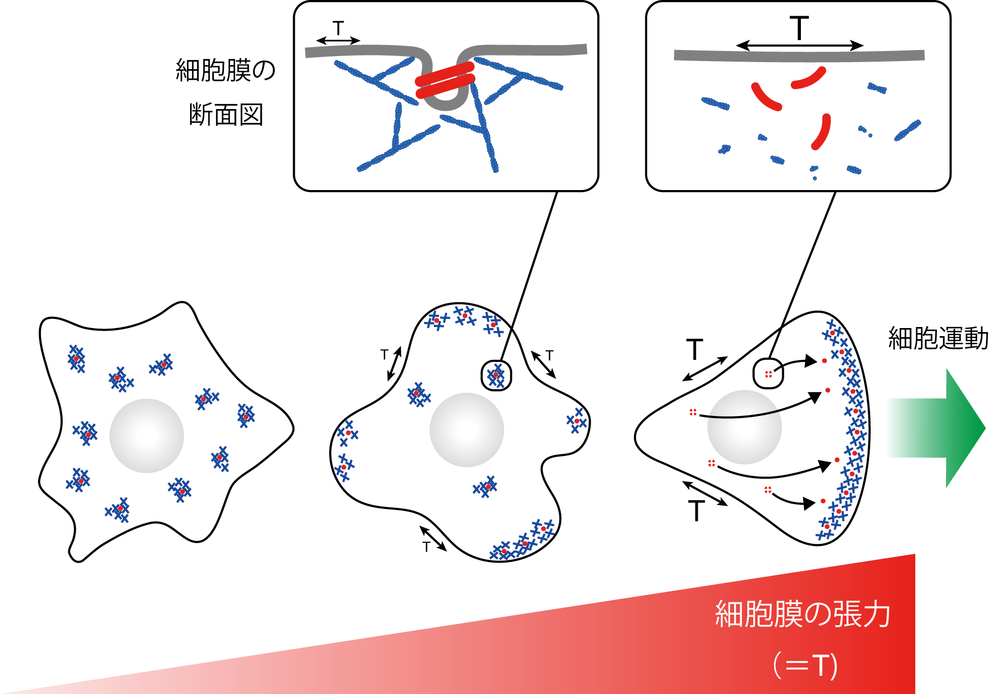
■ 研究シーズ- 細胞膜の形状と力学制御による疾患の理解シーズカテゴリ:ライフサイエンス研究キーワード:細胞膜, 細胞運動, 膜張力, がん細胞の浸潤転移, 細胞内情報伝達研究の背景と目的:「細胞膜」は細胞の内と外を隔てる境界であり、細胞を生命の基本単位として規定している。細胞膜は細胞内外の物質輸送と情報変換における重要な「反応の場」として機能するだけでなく、細胞の分裂や運動、分化などに伴いダイナミックな形態変化を遂げる柔軟な構造体である。細胞膜の動態は、膜を曲げるタンパク質と膜にかかる張力により規定されると考えられ、これらの因子による分子機構の解明を目指している。研究内容:細胞膜形状とアクチン細胞骨格をつなぐF-BARタンパク質ファミリーをはじめとする、数多くの膜制御因子を発見し (Science 2001; Dev. Cell 2005; Sci. Signal. 2009; J. Cell Biol. 2011)、F-BARタンパク質の細胞膜張力センサー分子としての機能を明らかにした (Nat. Cell Biol. 2015)。最近では、光ピンセットを用いて膜張力を計測することで、細胞膜にかかる張力がF-BARタンパク質に対抗し、悪性がん細胞の浸潤能を抑制することを見出している (Nat. Commun. 2021)。これらの研究を通じて、細胞膜張力がアクチン重合マシナリーとの相互フィードバックを介して、がん細胞浸潤のマスターレギュレーターとなるモデルの提示を行った。
 期待される効果や応用分野:がんを始めとする、細胞の分裂や移動を含むダイナミズムを基盤とする疾患は、これまで個別の分子の活性やこれに影響を与える遺伝子変異に着目したアプローチが中心であった。本研究は、より細胞の物性に近い「細胞膜の硬さ・柔らかさ」とアクチン重合に駆動される「力」という視点からこれらの疾患を捉え直す試みであり、これまでに存在しなかった画期的な創薬ターゲットが期待できる。
titoh people.kobe-u.ac.jp ※左記の「@」は画像ですので、ご利用の際はテキストの「@」を入力してください。
people.kobe-u.ac.jp ※左記の「@」は画像ですので、ご利用の際はテキストの「@」を入力してください。