SEARCH
検索詳細
木村 行宏大学院農学研究科 生命機能科学専攻准教授
研究者基本情報
■ 学位■ 研究ニュース
■ 研究キーワード
■ 研究分野
研究活動情報
■ 論文- Abstract Background Fermented katsuobushi, a traditional Japanese seasoning, is produced from skipjack tuna through smoking, drying and fermentation by xerophilic Aspergillus molds, primarily Aspergillus chevalieri and Aspergillus pseudoglaucus. In this study, we characterized lipolytic enzymes (cLip_1 to cLip_5 and pLip_1 to pLip_3) to clarify their roles in lipid hydrolysis during katsuobushi production under low water activity. Results The enzymes showed significant diversity in their activity, stability and substrate specificity, and in the hydrolysis profiles of their reactions with fish oil. Phylogenetic analyses revealed that cLip_5 showed a high identity with pLip_2 (94%) and these enzymes formed a phylogenetic cluster with filamentous fungal lipases. Purified recombinant enzymes (rcLip_1, rcLip_2, rcLip_4 and rcLip_5) and wild‐type enzymes (cLip_3 and pLip_3) showed varying substrate preferences toward p‐nitrophenyl esters. The addition of glycerol to reduce the water activity in the reaction mixture led to increased activities of rcLip_1 and rcLip_4, but it did not affect the activity of the other three enzymes. Among the tested six enzymes, cLip_5 showed the highest hydrolytic activity toward fish oil. The cLip_5 and pLip_2 gene transcript levels were moderately high in strains MK86 and MK88, respectively. Conclusion cLip_5 and its homolog pLip_2 were identified as the most promising enzymes for katsuobushi fermentation, because of their high hydrolytic activities toward fish oil and adaptability to low water activity conditions. These findings support the selection of optimal Aspergillus strains as starter cultures to potentially shorten the fermentation time and improve the quality and shelf life of katsuobushi. © 2025 Society of Chemical Industry.Wiley, 2025年02月, Journal of the Science of Food and Agriculture, 英語[査読有り]研究論文(学術雑誌)
- Springer Science and Business Media LLC, 2025年02月, Nature Communications, 16(1) (1), 英語[査読有り]研究論文(学術雑誌)
- Abstract Rhodothalassium (Rts.) salexigens is a halophilic purple nonsulfur bacterium and the sole species in the genus Rhodothalassium, which is itself the sole genus in the family Rhodothalassiaceae and sole family in the order Rhodothalassiales (class Alphaproteobacteria). The genome of this phylogenetically unique phototroph comprises 3.35 Mb and is highly chimeric, with nearly half of its genes originating from families other than the Rhodothalassiaceae, many of which lack phototrophic species. Photosynthesis genes in Rts. salexigens are not arranged in a typical photosynthesis gene cluster but are scattered across the genome, suggesting an origin from horizontal transfers. Despite an encoded RuBisCO, autotrophy has not been observed in Rts. salexigens, and enzymes that oxidize common inorganic electron donors are not encoded. Phospholipid biosynthesis in Rts. salexigens is restricted, and phosphoglycerolipids are the only phospholipids present in its intracytoplasmic membranes. Rts. salexigens fixes nitrogen using a Mo-containing nitrogenase and uses ammonia despite previous results that indicated it was a glutamate auxotroph. Glycine betaine is the sole osmolyte in Rts. salexigens, and enzymes are encoded that facilitate both its uptake and its biosynthesis from glycine. The genomic data also support chemotactic swimming motility, growth over a range of salinities, and the production of membrane-strengthening hopanoids.Springer Science and Business Media LLC, 2025年01月, Extremophiles, 29(1) (1)[査読有り]研究論文(学術雑誌)
- Springer Science and Business Media LLC, 2025年01月, Communications Biology, 8(1) (1), 英語[査読有り]研究論文(学術雑誌)
- Springer Science and Business Media LLC, 2024年12月, Communications Biology, 7(1) (1)[査読有り]研究論文(学術雑誌)
- American Chemical Society (ACS), 2024年12月, Biochemistry, 64(1) (1), 170 - 179, 英語[査読有り]研究論文(学術雑誌)
- Elsevier BV, 2024年08月, Biochimica et Biophysica Acta (BBA) - Bioenergetics, 149503 - 149503, 英語[査読有り]研究論文(学術雑誌)
- ABSTRACT Halorhodospira (Hlr.) halochloris is a triply extremophilic phototrophic purple sulfur bacterium, as it is thermophilic, alkaliphilic, and extremely halophilic. The light‐harvesting‐reaction center (LH1–RC) core complex of this bacterium displays an LH1‐Qy transition at 1,016 nm, which is the lowest‐energy wavelength absorption among all known phototrophs. Here we report the cryo‐EM structure of the LH1–RC at 2.42 Å resolution. The LH1 complex forms a tricyclic ring structure composed of 16 αβγ‐polypeptides and one αβ‐heterodimer around the RC. From the cryo‐EM density map, two previously unrecognized integral membrane proteins, referred to as protein G and protein Q, were identified. Both of these proteins are single transmembrane‐spanning helices located between the LH1 ring and the RC L‐subunit and are absent from the LH1–RC complexes of all other purple bacteria of which the structures have been determined so far. Besides bacteriochlorophyll b molecules (B1020) located on the periplasmic side of the Hlr. halochloris membrane, there are also two arrays of bacteriochlorophyll b molecules (B800 and B820) located on the cytoplasmic side. Only a single copy of a carotenoid (lycopene) was resolved in the Hlr. halochloris LH1–α3β3 and this was positioned within the complex. The potential quinone channel should be the space between the LH1–α3β3 that accommodates the single lycopene but does not contain a γ‐polypeptide, B800 and B820. Our results provide a structural explanation for the unusual Qy red shift and carotenoid absorption in the Hlr. halochloris spectrum and reveal new insights into photosynthetic mechanisms employed by a species that thrives under the harshest conditions of any phototrophic microorganism known.Wiley, 2024年02月, Journal of Integrative Plant Biology, 英語[査読有り]研究論文(学術雑誌)
- Abstract The ecological success of purple sulfur bacteria (PSB) is linked to their ability to collect near‐infrared solar energy by membrane‐integrated, pigment–protein photocomplexes. These include a Core complex containing both light‐harvesting 1 (LH1) and reaction centre (RC) components (called the LH1–RC photocomplex) present in all PSB and a peripheral light‐harvesting complex present in most but not all PSB. In research to explain the unusual absorption properties of the thermophilic purple sulfur bacterium Thermochromatium tepidum, Ca2+ was discovered bound to LH1 polypeptides in its LH1–RC; further work showed that calcium controls both the thermostability and unusual spectrum of the Core complex. Since then, Ca2+ has been found in the LH1–RC photocomplexes of several other PSB, including mesophilic species, but not in the LH1–RC of purple non‐sulfur bacteria. Here we focus on four species of PSB—two thermophilic and two mesophilic—and describe how Ca2+ is integrated into and affects their photosynthetic machinery and why this previously overlooked divalent metal is a key nutrient for their ecological success.Wiley, 2024年02月, Environmental Microbiology, 26(2) (2), 英語[査読有り][招待有り]研究論文(学術雑誌)
- Abstract The mesophilic purple sulfur phototrophic bacterium Allochromatium (Alc.) vinosum (bacterial family Chromatiaceae) has been a favored model for studies of bacterial photosynthesis and sulfur metabolism, and its core light-harvesting (LH1) complex has been a focus of numerous studies of photosynthetic light reactions. However, despite intense efforts, no high-resolution structure and thorough biochemical analysis of the Alc. vinosum LH1 complex have been reported. Here we present cryo-EM structures of the Alc. vinosum LH1 complex associated with reaction center (RC) at 2.24 Å resolution. The overall structure of the Alc. vinosum LH1 resembles that of its moderately thermophilic relative Alc. tepidum in that it contains multiple pigment-binding α- and β-polypeptides. Unexpectedly, however, six Ca ions were identified in the Alc. vinosum LH1 bound to certain α1/β1- or α1/β3-polypeptides through a different Ca2+-binding motif from that seen in Alc. tepidum and other Chromatiaceae that contain Ca2+-bound LH1 complexes. Two water molecules were identified as additional Ca2+-coordinating ligands. Based on these results, we reexamined biochemical and spectroscopic properties of the Alc. vinosum LH1–RC. While modest but distinct effects of Ca2+ were detected in the absorption spectrum of the Alc. vinosum LH1 complex, a marked decrease in thermostability of its LH1–RC complex was observed upon removal of Ca2+. The presence of Ca2+ in the photocomplex of Alc. vinosum suggests that Ca2+-binding to LH1 complexes may be a common adaptation in species of Chromatiaceae for conferring spectral and thermal flexibility on this key component of their photosynthetic machinery.Springer Science and Business Media LLC, 2024年02月, Communications Biology, 7(1) (1), 英語[査読有り]研究論文(学術雑誌)
- Elsevier BV, 2023年11月, Biochimica et Biophysica Acta (BBA) - Bioenergetics, 1864(4) (4), 149001 - 149001, 英語[査読有り]研究論文(学術雑誌)
- A salt-tolerant exo-ss-1,3-glucosidase (BGL_MK86) was cloned from the xerophilic mold Aspergillus chevalieri MK86 and heterologously expressed in A. oryzae. Phylogenetic analysis suggests that BGL_MK86 belongs to glycoside hydrolase family 5 (aryl- phospho-ss- D-glucosidase, BglC), and exhibits D-glucose tolerance. Recombinant BGL_MK86 (rBGL_MK86) exhibited 100-fold higher expression than native BGL_MK86. rBGL_MK86 was active over a wide range of NaCl concentrations [0%-18% (w/v)] and showed increased substrate affinity for p-nitrophenyl-ss-D-glucopyranoside (pNPBG) and turnover number (k(cat)) in the presence of NaCl. The enzyme was stable over a broad pH range (5.5-9.5). The optimum reaction pH and temperature for hydrolysis of pNPBG were 5.5 and 45 degrees C, respectively. rBGL_MK86 acted on the ss-1,3-linked glucose dimer laminaribiose, but not ss-1,4-linked or ss-1,6-linked glucose dimers (cellobiose or gentiobiose). It showed tenfold higher activity toward laminarin (a linear polymer of ss-1,3 glucan) from Laminaria digitata than laminarin (ss-1,3/ss-1,6 glucan) from Eisenia bicyclis, likely due to its inability to act on ss-1,6-linked glucose residues. The ss-glucosidase retained hydrolytic activity toward crude laminarin preparations from marine biomass in moderately high salt concentrations. These properties indicate wide potential applications of this enzyme in saccharification of salt-bearing marine biomass.SPRINGER, 2023年09月, ARCHIVES OF MICROBIOLOGY, 205(9) (9), 英語[査読有り]研究論文(学術雑誌)
- Although several species of purple sulfur bacteria inhabit soda lakes, Rhodobaca bogoriensis is the first purple nonsulfur bacterium cultured from such highly alkaline environments. Rhodobaca bogoriensis strain LBB1(T) was isolated from Lake Bogoria, a soda lake in the African Rift Valley. The phenotype of Rhodobaca bogoriensis is unique among purple bacteria; the organism is alkaliphilic but not halophilic, produces carotenoids absent from other purple nonsulfur bacteria, and is unable to grow autotrophically or fix molecular nitrogen. Here we analyze the draft genome sequence of Rhodobaca bogoriensis to gain further insight into the biology of this extremophilic purple bacterium. The strain LBB1(T) genome consists of 3.91 Mbp with no plasmids. The genome sequence supports the defining characteristics of strain LBB1(T), including its (1) production of a light-harvesting 1-reaction center (LH1-RC) complex but lack of a peripheral (LH2) complex, (2) ability to synthesize unusual carotenoids, (3) capacity for both phototrophic (anoxic/light) and chemotrophic (oxic/dark) energy metabolisms, (4) utilization of a wide variety of organic compounds (including acetate in the absence of a glyoxylate cycle), (5) ability to oxidize both sulfide and thiosulfate despite lacking the capacity for autotrophic growth, and (6) absence of a functional nitrogen-fixation system for diazotrophic growth. The assortment of properties in Rhodobaca bogoriensis has no precedent among phototrophic purple bacteria, and the results are discussed in relation to the organism's soda lake habitat and evolutionary history.SPRINGER JAPAN KK, 2023年08月, EXTREMOPHILES, 27(2) (2), 英語[査読有り]研究論文(学術雑誌)
- Dried bonito broth, made from katsuobushi flakes, is a commonly used seasoning in Japanese cuisine. While extracting the broth, high-quality proteins are left behind in the katsuobushi grounds, which could be a valuable source of bioactive substances via a hydrolysis process. The xerophilic Aspergillus sydowii showed potential for protein hydrolysis and production of amino acids and soluble peptides through solid-state fermentation. Several peptidases involved in katsuobushi protein hydrolysis were purified from the crude enzyme preparations and identified as aminopeptidase, trypsin-like serine protease, and elastinolytic serine protease. The hydrolysates from the culture contained glutamic acid, leucine, and lysine and markedly increased soluble peptides with molecular weights ranging from 0.2 to 17 kDa. Electronic tongue analysis indicated that the hydrolysates had substantially stronger salty and umami tastes. Moreover, the hydrolysates exhibited radical-scavenging activities, ferric ion reducing antioxidant power, and angiotensin converting enzyme inhibitory activity, as well as pro-moting growth in lactic acid bacteria. In addition, de novo peptide sequencing was performed for peptides fractionated from the hydrolysates, and eight peptides were identified. The hydrolysates produced by A. sydowii fermentation could have potential applications in the food industry as natural flavor enhancers and functional ingredients with several bioactivities.ELSEVIER SCI LTD, 2023年07月, PROCESS BIOCHEMISTRY, 130, 534 - 544, 英語[査読有り]研究論文(学術雑誌)
- Elsevier BV, 2023年04月, Enzyme and Microbial Technology, 110240 - 110240, 英語[査読有り]研究論文(学術雑誌)
- Structural information on the circular arrangements of repeating pigment-polypeptide subunits in antenna proteins of purple photosynthetic bacteria is a clue to a better understanding of molecular mechanisms for the ring-structure formation and efficient light harvesting of such antennas. Here, we have analyzed the ring structure of light-harvesting complex 2 (LH2) from the thermophilic purple bacterium Thermochromatium tepidum (tepidum-LH2) by atomic force microscopy. The circular arrangement of the tepidum-LH2 subunits was successfully visualized in a lipid bilayer. The average top-to-top distance of the ring structure, which is correlated with the ring size, was 4.8 ± 0.3 nm. This value was close to the top-to-top distance of the octameric LH2 from Phaeospirillum molischianum (molischianum-LH2) by the previous analysis. Gaussian distribution of the angles of the segments consisting of neighboring subunits in the ring structures of tepidum-LH2 yielded a median of 44°, which corresponds to the angle for the octameric circular arrangement (45°). These results indicate that tepidum-LH2 has a ring structure consisting of eight repeating subunits. The coincidence of an octameric ring structure of tepidum-LH2 with that of molischianum-LH2 is consistent with the homology of amino acid sequences of the polypeptides between tepidum-LH2 and molischianum-LH2.2023年03月, Photosynthesis research, 英語, 国際誌[査読有り]研究論文(学術雑誌)
- Rhodobacter (Rba.) capsulatus has been a favored model for studies of all aspects of bacterial photosynthesis. This purple phototroph contains PufX, a polypeptide crucial for dimerization of the light-harvesting 1-reaction center (LH1-RC) complex, but lacks protein-U, a U-shaped polypeptide in the LH1-RC of its close relative Rba. sphaeroides. Here we present a cryo-EM structure of the Rba. capsulatus LH1-RC purified by DEAE chromatography. The crescent-shaped LH1-RC exhibits a compact structure containing only 10 LH1 αβ-subunits. Four αβ-subunits corresponding to those adjacent to protein-U in Rba. sphaeroides were absent. PufX in Rba. capsulatus exhibits a unique conformation in its N-terminus that self-associates with amino acids in its own transmembrane domain and interacts with nearby polypeptides, preventing it from interacting with proteins in other complexes and forming dimeric structures. These features are discussed in relation to the minimal requirements for the formation of LH1-RC monomers and dimers, the spectroscopic behavior of both the LH1 and RC, and the bioenergetics of energy transfer from LH1 to the RC.2023年02月, Nature communications, 14(1) (1), 846 - 846, 英語, 国際誌[査読有り]研究論文(学術雑誌)
- American Chemical Society (ACS), 2023年01月, The Journal of Physical Chemistry B, 英語[査読有り][招待有り]研究論文(学術雑誌)
- Abstract Rhodobacter sphaeroides is a model organism in bacterial photosynthesis, and its light-harvesting-reaction center (LH1–RC) complex contains both dimeric and monomeric forms. Here we present cryo-EM structures of the native LH1–RC dimer and an LH1–RC monomer lacking protein-U (ΔU). The native dimer reveals several asymmetric features including the arrangement of its two monomeric components, the structural integrity of protein-U, the overall organization of LH1, and rigidities of the proteins and pigments. PufX plays a critical role in connecting the two monomers in a dimer, with one PufX interacting at its N-terminus with another PufX and an LH1 β-polypeptide in the other monomer. One protein-U was only partially resolved in the dimeric structure, signaling different degrees of disorder in the two monomers. The ΔU LH1–RC monomer was half-moon-shaped and contained 11 α- and 10 β-polypeptides, indicating a critical role for protein-U in controlling the number of αβ-subunits required for dimer assembly and stabilization. These features are discussed in relation to membrane topology and an assembly model proposed for the native dimeric complex.Springer Science and Business Media LLC, 2022年12月, Nature Communications, 13(1) (1), 1904 - 1904, 英語, 国際誌[査読有り]研究論文(学術雑誌)
- Abstract Rhodopila globiformis is the most acidophilic of anaerobic purple phototrophs, growing optimally in culture at pH 5. Here we present a cryo-EM structure of the light-harvesting 1–reaction center (LH1–RC) complex from Rhodopila globiformis at 2.24 Å resolution. All purple bacterial cytochrome (Cyt, encoded by the gene pufC) subunit-associated RCs with known structures have their N-termini truncated. By contrast, the Rhodopila globiformis RC contains a full-length tetra-heme Cyt with its N-terminus embedded in the membrane forming an α-helix as the membrane anchor. Comparison of the N-terminal regions of the Cyt with PufX polypeptides widely distributed in Rhodobacter species reveals significant structural similarities, supporting a longstanding hypothesis that PufX is phylogenetically related to the N-terminus of the RC-bound Cyt subunit and that a common ancestor of phototrophic Proteobacteria contained a full-length tetra-heme Cyt subunit that evolved independently through partial deletions of its pufC gene. Eleven copies of a novel γ-like polypeptide were also identified in the bacteriochlorophyll a-containing Rhodopila globiformis LH1 complex; γ-polypeptides have previously been found only in the LH1 of bacteriochlorophyll b-containing species. These features are discussed in relation to their predicted functions of stabilizing the LH1 structure and regulating quinone transport under the warm acidic conditions.Springer Science and Business Media LLC, 2022年11月, Communications Biology, 5(1) (1)[査読有り]研究論文(学術雑誌)
- Springer Science and Business Media LLC, 2022年10月, 3 Biotech, 12(10) (10)[査読有り]研究論文(学術雑誌)
- Halorhodospira (Hlr.) species are the most halophilic and alkaliphilic of all purple bacteria. Hlr. halochloris exhibits the lowest LH1 Qy transition energy among phototrophic organisms and is the only known triply extremophilic anoxygenic phototroph, displaying a thermophilic, halophilic, and alkaliphilic phenotype. Recently, we reported that electrostatic charges are responsible for the unusual spectroscopic properties of the Hlr. halochloris LH1 complex. In the present work, we examined the effects of salt and pH on the spectroscopic properties and thermal stability of LH1-RCs from Hlr. halochloris compared with its mesophilic counterpart, Hlr. abdelmalekii. Experiments in which the photocomplexes were subjected to different levels of salt or variable pH revealed that the thermal stability of LH1-RCs from both species was largely retained in the presence of high salt concentrations and/or at alkaline pH but was markedly reduced by lowering the salt concentration and/or pH. Based on the amino acid sequences of LH1 polypeptides and their composition of acidic/basic residues and the Hofmeister series for cation/anion species, we discuss the importance of electrostatic charge in stabilizing the Hlr. halochloris LH1-RC complex to allow it to perform photosynthesis in its warm, hypersaline, and alkaline habitat.MDPI AG, 2022年05月, Microorganisms, 10(5) (5), 959 - 959, 英語[査読有り][招待有り]研究論文(学術雑誌)
- The mildly thermophilic purple phototrophic bacterium Allochromatium tepidum provides a unique model for investigating various intermediate phenotypes observed between those of thermophilic and mesophilic counterparts. The core light-harvesting (LH1) complex from A. tepidum exhibits an absorption maximum at 890 nm and mildly enhanced thermostability, both of which are Ca2+-dependent. However, it is unknown what structural determinants might contribute to these properties. Here, we present a cryo-EM structure of the reaction center-associated LH1 complex at 2.81 Å resolution, in which we identify multiple pigment-binding α- and β-polypeptides within an LH1 ring. Of the 16 α-polypeptides, we show that six (α1) bind Ca2+ along with β1- or β3-polypeptides to form the Ca2+-binding sites. This structure differs from that of fully Ca2+-bound LH1 from Thermochromatium tepidum, enabling determination of the minimum structural requirements for Ca2+-binding. We also identified three amino acids (Trp44, Asp47, and Ile49) in the C-terminal region of the A. tepidum α1-polypeptide that ligate each Ca ion, forming a Ca2+-binding WxxDxI motif that is conserved in all Ca2+-bound LH1 α-polypeptides from other species with reported structures. The partial Ca2+-bound structure further explains the unusual phenotypic properties observed for this bacterium in terms of its Ca2+-requirements for thermostability, spectroscopy, and phototrophic growth, and supports the hypothesis that A. tepidum may represent a "transitional" species between mesophilic and thermophilic purple sulfur bacteria. The characteristic arrangement of multiple αβ-polypeptides also suggests a mechanism of molecular recognition in the expression and/or assembly of the LH1 complex that could be regulated through interactions with reaction center subunits.Elsevier BV, 2022年04月, Journal of Biological Chemistry, 298(6) (6), 101967 - 101967, 英語, 国際誌[査読有り]研究論文(学術雑誌)
- AIP Publishing, 2022年02月, The Journal of Chemical Physics[査読有り][招待有り]研究論文(学術雑誌)
- Wiley, 2022年02月, Journal of Basic Microbiology, 62(2) (2), 174 - 184[査読有り]研究論文(学術雑誌)
- The interactions of chlorophyll (Chl) and bacteriochlorophyll (BChl) pigments with the polypeptides in photosynthetic light-harvesting proteins are responsible for controlling the absorption energy of (B)Chls in protein matrixes. The binding pocket of B800 BChl a in LH2 proteins, which are peripheral light-harvesting proteins in purple photosynthetic bacteria, is useful for studying such structure-property relationships. We report the reconstitution of Chl f, which has the formyl group at the 2-position, in the B800 cavity of LH2 from the purple bacterium Rhodoblastus acidophilus. The Q(y) absorption band of Chl f in the B800 cavity was shifted by 14 nm to longer wavelength compared to that of the corresponding five-coordinated monomer in acetone. This redshift was larger than that of Chl a and Chl b. Resonance Raman spectroscopy indicated hydrogen bonding between the 2-formyl group of Chl f and the LH2 polypeptide. These results suggest that this hydrogen bonding contributes to the Q(y) redshift of Chl f. Furthermore, the Q(y) redshift of Chl f in the B800 cavity was smaller than that of Chl d. This may have arisen from the different patterns of hydrogen bonding between Chl f and Chl d and/or from the steric hindrance of the 3-vinyl group in Chl f.Wiley, 2022年01月, Photochemistry and Photobiology, 98(1) (1), 169 - 174, 英語[査読有り]研究論文(学術雑誌)
- Springer Science and Business Media LLC, 2021年12月, Nature Communications, 12(1) (1), 6300 - 6300, 英語, 国際誌
Abstract Rhodobacter (Rba .)sphaeroides is the most widely used model organism in bacterial photosynthesis. The light-harvesting-reaction center (LH1-RC) core complex of this purple phototroph is characterized by the co-existence of monomeric and dimeric forms, the presence of the protein PufX, and approximately two carotenoids per LH1 αβ-polypeptides. Despite many efforts, structures of theRba. sphaeroides LH1-RC have not been obtained at high resolutions. Here we report a cryo-EM structure of the monomeric LH1-RC fromRba. sphaeroides strain IL106 at 2.9 Å resolution. The LH1 complex forms a C-shaped structure composed of 14 αβ-polypeptides around the RC with a large ring opening. From the cryo-EM density map, a previously unrecognized integral membrane protein, referred to as protein-U, was identified. Protein-U has a U-shaped conformation near the LH1-ring opening and was annotated as a hypothetical protein in theRba. sphaeroides genome. Deletion of protein-U resulted in a mutant strain that expressed a much-reduced amount of the dimeric LH1-RC, indicating an important role for protein-U in dimerization of the LH1-RC complex. PufX was located opposite protein-U on the LH1-ring opening, and both its position and conformation differed from that of previous reports of dimeric LH1-RC structures obtained at low-resolution. Twenty-six molecules of the carotenoid spheroidene arranged in two distinct configurations were resolved in theRba. sphaeroides LH1 and were positioned within the complex to block its channels. Our findings offer an exciting new view of the core photocomplex ofRba. sphaeroides and the connections between structure and function in bacterial photocomplexes in general.[査読有り]研究論文(学術雑誌) - Springer Science and Business Media LLC, 2021年12月, Nature Communications, 12(1) (1), 英語
Abstract Photosynthetic electron transfers occur through multiple components ranging from small soluble proteins to large integral membrane protein complexes. Co-crystallization of a bacterial photosynthetic electron transfer complex that employs weak hydrophobic interactions was achieved by using high-molar-ratio mixtures of a soluble donor protein (high-potential iron-sulfur protein, HiPIP) with a membrane-embedded acceptor protein (reaction center, RC) at acidic pH. The structure of the co-complex offers a snapshot of a transient bioenergetic event and revealed a molecular basis for thermodynamically unfavorable interprotein electron tunneling. HiPIP binds to the surface of the tetraheme cytochrome subunit in the light-harvesting (LH1) complex-associated RC in close proximity to the low-potential heme-1 group. The binding interface between the two proteins is primarily formed by uncharged residues and is characterized by hydrophobic features. This co-crystal structure provides a model for the detailed study of long-range trans-protein electron tunneling pathways in biological systems.[査読有り]研究論文(学術雑誌) - Halorhodospira (Hlr.) halochloris is a unique phototrophic purple bacterium because it is a triple extremophile-the organism is thermophilic, alkalophilic, and halophilic. The most striking photosynthetic feature of Hlr. halochloris is that the bacteriochlorophyll (BChl) b-containing core light-harvesting (LH1) complex surrounding its reaction center (RC) exhibits its LH1 Q(y) absorption maximum at 1016 nm, which is the lowest transition energy among phototrophic organisms. Here we report that this extraordinarily red-shifted LH1 Q(y) band of Hlr. halochloris exhibits interconvertible spectral shifts depending on the electrostatic charge distribution around the BChl b molecules. The 1016 nm band of the Hlr. halochloris LH1-RC complex was blue-shifted to 958 nm upon desalting or pH decrease but returned to its original position when supplemented with salts or pH increase. Resonance Raman analysis demonstrated that these interconvertible spectral shifts are not associated with the strength of hydrogen-bonding interactions between BChl b and LH1 polypeptides. Furthermore, circular dichroism signals for the LH1 Q(y) transition of Hlr. halochloris appeared with a positive sign (as in BChl b-containing Blastochloris species) and opposite those of BChl a-containing purple bacteria, possibly due to a combined effect of slight differences in the transition dipole moments between BChl a and BChl b and in the interactions between adjacent BChls in their assembled state. Based on these findings and LH1 amino acid sequences, it is proposed that Hlr. halochloris evolved its unique and tunable light-harvesting system with electrostatic charges in order to carry out photosynthesis and thrive in its punishing hypersaline and alkaline habitat.ELSEVIER, 2021年11月, BIOCHIMICA ET BIOPHYSICA ACTA-BIOENERGETICS, 1862(11) (11), 英語[査読有り]研究論文(学術雑誌)
- Elsevier BV, 2021年09月, International Journal of Food Microbiology, 353, 109299 - 109299[査読有り]研究論文(学術雑誌)
- Light-harvesting complex 1 (LH1) of the thermophilic purple sulfur bacterium Thermochromatium tepidum can be expressed in the purple non-sulfur bacterium Rhodobacter sphaeroides and forms a functional RC-LH1 complex with the native Rba. sphaeroides reaction center (Nagashima, K. V. P., et al. Proc. Natl. Acad. Sci. U. S. A. 2017, 114, 10906-10911). Although there is a large uphill energy gap between Tch. tepidum LH1 and the Rba. sphaeroides RC in this chimeric complex, it has been shown that light energy can be transferred, consistent with that seen in the native Rba. sphaeroides RC-LH1 complex. In this study, the contribution of this chimeric complex to growth and photosynthetic energy conversion in the hybrid organism was quantified. The mutant synthesizing this chimeric complex was grown phototrophically under 940 nm light-emitting diode (LED) light preferentially absorbed by Tch. tepidum LH1 and showed faster growth at low intensities of this wavelength than both a mutant strain of Rba. sphaeroides lacking LH2 and a mutant lacking all light-harvesting complexes. When grown with 850 nm LED light, the strain containing the native Rba. sphaeroides LH1-RC grew faster than the chimeric strain. Electron transfer from the RC to the membrane-integrated cytochrome bc(1) complex was also estimated by flash-induced absorption changes in heme b. The rate of ubiquinone transport through the LH1 ring structure in the chimeric strain was virtually the same as that in native Rba. sphaeroides. We conclude that Tch. tepidum LH1 can perform the physiological functions of native LH1 in Rba. sphaeroides.AMER CHEMICAL SOC, 2021年09月, BIOCHEMISTRY, 60(36) (36), 2685 - 2690, 英語[査読有り]研究論文(学術雑誌)
- Rhodospirillum (Rsp.) rubrum is one of the most widely used model organisms in bacterial photosynthesis. This purple phototroph is characterized by the presence of both rhodoquinone (RQ) and ubiquinone as electron carriers and bacteriochlorophyll (BChl) a esterified at the propionic acid side chain by geranylgeraniol (BChl a(G)) instead of phytol. Despite intensive efforts, the structure of the light-harvesting-reaction center (LH1-RC) core complex from Rsp. rubrum remains at low resolutions. Using cryo-EM, here we present a robust new view of the Rsp. rubrum LH1-RC at 2.76 angstrom resolution. The LH1 complex forms a closed, slightly elliptical ring structure with 16 alpha beta-polypeptides surrounding the RC. Our biochemical analysis detected RQ molecules in the purified LH1-RC, and the cryo-EM density map specifically positions RQ at the Q(A) site in the RC. The geranylgeraniol side chains of BChl a(G) coordinated by LH1 beta-polypeptides exhibit a highly homologous tail-up conformation that allows for interactions with the bacteriochlorin rings of nearby LH1 alpha-associated BChls a(G). The structure also revealed key protein-protein interactions in both Nand C-terminal regions of the LH1 alpha beta-polypeptides, mainly within a face-to-face structural subunit. Our high-resolution Rsp. rubrum LH1-RC structure provides new insight for evaluating past experimental and computational results obtained with this old organism over many decades and lays the foundation for more detailed exploration of light-energy conversion, quinone transport, and structure-function relationships in this pigment-protein complex.AMER CHEMICAL SOC, 2021年08月, BIOCHEMISTRY, 60(32) (32), 2483 - 2491, 英語[査読有り]研究論文(学術雑誌)
- Cold Spring Harbor Laboratory, 2021年06月, 英語
Abstract We present a cryo-EM structure of the monomeric light-harvesting-reaction center (LH1-RC) core complex from photosynthetic purple bacteriumRhodobacter (Rba .)sphaeroides at 2.9 Å resolution. The LH1 complex forms a C-shaped structure composed of 14 αβ-polypeptides around the RC with a large ring opening. From the cryo-EM density map, a previously unrecognized integral membrane protein, referred to as protein-U, was identified. Protein-U has a U-shaped conformation near the LH1-ring opening and was annotated as a hypothetical protein in theRba. sphaeroides genome. Deletion of protein-U resulted in a mutant strain that expressed a much-reduced amount of the dimeric LH1-RC, indicating an important role for protein-U in dimerization of the LH1-RC complex. PufX was located opposite protein-U on the LH1-ring opening, and both its position and conformation differed from that of previous reports of dimeric LH1-RC structures obtained at low-resolution. Twenty-six molecules of the carotenoid spheroidene arranged in two distinct configurations were resolved in theRba. sphaeroides LH1 and were positioned within the complex to block its pores. Our findings offer a new view of the core photocomplex ofRba. sphaeroides and the connections between structure and function in bacterial photocomplexes in general.研究論文(学術雑誌) - Cold Spring Harbor Laboratory, 2021年05月
Abstract We present a cryo-EM structure of the light-harvesting-reaction center (LH1-RC) core complex from purple phototrophic bacteriumRhodospirillum (Rsp .)rubrum at 2.76 Å resolution. The LH1 complex forms a closed, slightly elliptical ring structure with 16 αβ-polypeptides surrounding the RC. Our biochemical analysis detected rhodoquinone (RQ) molecules in the purified LH1-RC, and the cryo-EM density map specifically positions RQ at the QA site in the RC. The geranylgeraniol sidechains of bacteriochlorophyll (BChl)a G coordinated by LH1 β-polypeptides exhibit a highly homologous tail-up conformation that allows for interactions with the bacteriochlorin rings of nearby LH1 α-associated BChlsa G. The structure also revealed key protein–protein interactions in both N- and C-terminal regions of the LH1 αβ-polypeptides, mainly within a face-to-face structural subunit. Our findings enable to evaluate past experimental and computational results obtained with this widely used organism and provide crucial information for more detailed exploration of light-energy conversion, quinone transport, and structure–function relationships in pigment-protein complexes. - Springer Science and Business Media LLC, 2021年05月, Photosynthesis Research, 148(1-2) (1-2), 77 - 86[査読有り]研究論文(学術雑誌)
- Control of the spectral overlap between energy donors and acceptors provides insight into excitation energy transfer (EET) mechanisms in photosynthetic light-harvesting proteins. Substitution of energy-donating B800 bacteriochlorophyll (BChl) a with other pigments in the light-harvesting complex 2 (LH2) of purple photosynthetic bacteria has been extensively performed; however, most studies on the B800 substitution have focused on the decrease in the spectral overlap integral with energy-accepting B850 BChl a by reconstitution of chlorophylls into the B800 site. Here, we reconstitute BChl b into the B800 site of the LH2 protein from Rhodoblastus acidophilus to increase the spectral overlap with B850 BChl a. BChl b in the B800 site had essentially the same hydrogen-bonding pattern as B800 BChl a, whereas it showed a red-shifted Q(y) absorption band at 831 nm. The EET rate from BChl b to B850 BChl a in the reconstituted LH2 was similar to that of native LH2 despite the red shift of the Q(y) band of the energy donor. These results demonstrate the importance of the contribution of the density of excitation states of the B850 circular assembly, which incorporates higher lying optically forbidden states, to intracomplex EET in LH2.American Chemical Society (ACS), 2021年03月, The Journal of Physical Chemistry B, 125(8) (8), 2009 - 2017, 英語[査読有り]研究論文(学術雑誌)
- Redox-active quinones play essential roles in efficient light energy conversion in type-II reaction centers of purple phototrophic bacteria. In the light-harvesting 1 reaction center (LH1-RC) complex of purple bacteria, Q(B) is converted to Q(B)H(2) upon light-induced reduction and Q(B)H(2) is transported to the quinone pool in the membrane through the LH1 ring. In the purple bacterium Rhodobacter sphaeroides, the C-shaped LH1 ring contains a gap for quinone transport. In contrast, the thermophilic purple bacterium Thermochromatium (Tch.) tepidum has a closed O-shaped LH1 ring that lacks a gap, and hence the mechanism of photosynthetic quinone transport is unclear. Here we detected light-induced Fourier transform infrared (FTIR) signals responsible for changes of Q(B) and its binding site that accompany photosynthetic quinone reduction in Tch. tepidum and characterized Q(B) and Q(B)H(2) marker bands based on their N-15- and C-13-isotopic shifts. Quinone exchanges were monitored using reconstituted photosynthetic membranes comprised of solubilized photosynthetic proteins, membrane lipids, and exogenous ubiquinone (UQ) molecules. In combination with C-13-labeling of the LH1-RC and replacement of native UQ(8) by ubiquinones of different tail lengths, we demonstrated that quinone exchanges occur efficiently within the hydrophobic environment of the lipid membrane and depend on the side chain length of UQ. These results strongly indicate that unlike the process in Rba. sphaeroides, quinone transport in Tch. tepidum occurs through the size-restricted hydrophobic channels in the closed LH1 ring and are consistent with structural studies that have revealed narrow hydrophobic channels in the Tch. tepidum LH1 transmembrane region.Elsevier BV, 2021年01月, Biochimica et Biophysica Acta (BBA) - Bioenergetics, 1862(1) (1), 148307 - 148307, 英語[査読有り]研究論文(学術雑誌)
- Springer Science and Business Media LLC, 2020年12月, Nature Communications, 11(1) (1), 4955 - 4955, 英語, 国際誌
Abstract The light-harvesting-reaction center complex (LH1-RC) from the purple phototrophic bacteriumThiorhodovibrio strain 970 exhibits an LH1 absorption maximum at 960 nm, the most red-shifted absorption for any bacteriochlorophyll (BChl)a -containing species. Here we present a cryo-EM structure of the strain 970 LH1-RC complex at 2.82 Å resolution. The LH1 forms a closed ring structure composed of sixteen pairs of the αβ-polypeptides. Sixteen Ca ions are present in the LH1 C-terminal domain and are coordinated by residues from the αβ-polypeptides that are hydrogen-bonded to BChla . The Ca2+-facilitated hydrogen-bonding network forms the structural basis of the unusual LH1 redshift. The structure also revealed the arrangement of multiple forms of α- and β-polypeptides in an individual LH1 ring. Such organization indicates a mechanism of interplay between the expression and assembly of the LH1 complex that is regulated through interactions with the RC subunits inside.[査読有り]研究論文(学術雑誌) - A soup stock made from katsuobushi is an important element of, and the basic seasoning responsible for the taste of, traditional Japanese cuisine. Fermented and ripened katsuobushi, called karebushi, is manufactured via a repeated molding process on the katsuobushi surface. Our aim was to characterize the surface Aspergillus community and their enzymes involved in the fermentation and ripening. Five dominant Aspergillus species isolated from the karebushi surface were identified-A. amstelodami, A. chevalieri, A. pseudoglaucus, A. ruber, and A. sydowii. Analyses were performed on final molding stage-samples from different manufacturers, and 1st to 4th molding stage-samples from the same manufacturer. The composition ratios of the five Aspergillus spp. varied according to the manufacturer of the karebushi. A. amstelodami and A. chevalieri tended to be detected as dominant species when the water content of the karebushi fillet was >15% and the fat content was >3.5%, respectively. In samples from a given manufacturer, the dominant species in the final molding stage tended to be A. chevalieri and A. pseudoglaucus. Mixed molds were cultured by solid-state fermentation using katsuobushi powder medium at two different water activity (aw) levels. Crude extracts from each culture showed lipase, aminopeptidase, carboxypeptidase, and protease activities. Notably, the crude extracts cultivated at 0.85 aw showed higher protease activity toward hemoglobin and lipase activity toward p-nitrophenyl palmitate than those at 0.95 aw. These hydrolytic enzymes are probably involved in decolorization of katsuobushi and lipid degradation during the long fermentative and ripening period. In addition, mixed cultures could transform 2,6-dimethoxyphenol into 1,2,3-trimethoxybenzene, previously reported as an attractive and mild flavor component. Our results may help promote the use of desirable Aspergillus spp. as starter cultures for manufacturers to stabilize and improve the quality of fermented and ripened karebushi.Elsevier BV, 2020年08月, International Journal of Food Microbiology, 327, 108654 - 108654, 英語, 国際誌[査読有り]研究論文(学術雑誌)
- Blastochloris tepida is a newly described thermophilic purple bacterium containing bacteriochlorophyll b. Using purified light-harvesting 1 reaction center (LH1-RC) core complexes from Blc. tepida, we compared the biochemical, spectroscopic, and thermal denaturation properties of these complexes with those of its mesophilic counterpart, Blc. viridis. Besides their growth temperature optima, a striking difference between the two species was seen in the carotenoid composition of their LH1-RC complexes. The more thermostable Blc. tepida complex contained more carotenoids with longer conjugation lengths (n > 9), such as lycopenes (n = 11), and had a total carotenoid content significantly higher than that of the Blc. viridis complex, irrespective of the light intensity used for growth. The thermostability of LH1-RCs from both Blc. tepida and Blc. viridis decreased significantly in cells grown in the presence of diphenylamine, a compound that inhibits the formation of highly conjugated carotenoids. In contrast to the thermophilic purple bacterium Thermochromatium tepidum, where Ca2+ is essential for LH1-RC thermostability, Ca2+ neither was present in nor had any effect on the thermostability of the Blc. tepida LH1 RC. These results point to a mechanism that carotenoids with elongated conjugations enhance hydrophobic interactions with proteins in the Blc. tepida LH1-RC, thereby allowing the complexes to withstand thermal denaturation. This conclusion is bolstered by a structural model of the Blc. tepida LH 1-RC and is the first example of photocomplex thermostability being linked to a carotenoid-based mechanism.AMER CHEMICAL SOC, 2020年06月, BIOCHEMISTRY, 59(25) (25), 2351 - 2358, 英語[査読有り]研究論文(学術雑誌)
- The manipulation of B800 bacteriochlorophyll (BChl) a in light-harvesting complex 2 (LH2) from the purple photosynthetic bacterium Phaeospirillum molischianum (molischianum-LH2) provides insight for understanding the energy transfer mechanism and the binding of cyclic tetrapyrroles in LH2 proteins since molischianum-LH2 is one of the two LH2 proteins whose atomic-resolution structures have been determined and is a representative of type-2 LH2 proteins. However, there is no report on the substitution of B800 BChl a in molischianum-LH2. We report the reconstitution of 3-acetyl chlorophyll (AcChl) a, which has a 17,18-dihydroporphyrin skeleton, to the B800 site in molischianum-LH2. The 3-acetyl group in AcChl a formed a hydrogen bond with beta'-Thr23 in essentially the same manner as native B800 BChl a, but this hydrogen bond was weaker than that of B800 BChl a. This change can be rationalized by invoking a small distortion in the orientation of the 3-acetyl group in the B800 cavity by dehydrogenation in the B-ring from BChl a. The energy transfer from AcChl a in the B800 site to B850 BChl a was about 5-fold slower than that from native B800 BChl a by a decrease of the spectral overlap between energy-donating AcChl a and energy-accepting B850 BChl a.AMER CHEMICAL SOC, 2020年03月, ACS OMEGA, 5(12) (12), 6817 - 6825, 英語[査読有り]研究論文(学術雑誌)
- Elsevier BV, 2019年10月, PROCESS BIOCHEMISTRY, 85, 156 - 163[査読有り]研究論文(学術雑誌)
- The light-harvesting 1 reaction center (LH1-RC) complex in the purple sulfur bacterium Thiorhodovibrio (Trv.) strain 970 cells exhibits its LH1 Q(y) transition at 973 nm, the lowest-energy Q(y) absorption among purple bacteria containing bacteriochlorophyll a (BChl a). Here we characterize the origin of this extremely red-shifted Qy transition. Growth of Try. strain 970 did not occur in cultures free of Ca2+, and elemental analysis of Ca2+-grown cells confirmed that purified Try. strain 970 LH1-RC complexes contained Ca2+. The LH1 Qy band of Try. strain 970 was blue-shifted from 959 to 875 nm upon Ca2+ depletion, but the original spectral properties were restored upon Ca2+ reconstitution, which also occurs with the thermophilic purple bacterium Thermochromatium (Tch.) tepidum. The amino acid sequences of the LH1 alpha- and beta-polypeptides from Try. strain 970 closely resemble those of Tch. tepidum; however, Ca2+ binding in the Try. strain 970 LH1-RC occurred more selectively than in Tch. tepidum LH1-RC and with a reduced affinity. Ultraviolet resonance Raman analysis indicated that the number of hydrogen-bonding interactions between BChl a and LH1 proteins of Try. strain 970 was significantly greater than for Tch. tepidum and that Ca2+ was indispensable for maintaining these bonds. Furthermore, perfusion-induced Fourier transform infrared analyses detected Ca2+-induced conformational changes in the binding site closely related to the unique spectral properties of Try. strain 970. Collectively, our results reveal an ecological strategy employed by Try. strain 970 of integrating Ca2+ into its LH1-RC complex to extend its light-harvesting capacity to regions of the near-infrared spectrum unused by other purple bacteria.AMER CHEMICAL SOC, 2019年06月, BIOCHEMISTRY, 58(25) (25), 2844 - 2852, 英語[査読有り]研究論文(学術雑誌)
- Elsevier BV, 2019年04月, PROCESS BIOCHEMISTRY, 79, 74 - 80[査読有り]研究論文(学術雑誌)
- 2019年04月, Biochimica Biophysica Acta, 1860, 461 - 468Phospholipid distributions in purple phototrophic bacteria and LH1-RC core complexes[査読有り]
- Engineering chlorophyll (Chl) pigments that are bound to photosynthetic light-harvesting proteins is one promising strategy to regulate spectral coverage for photon capture and to improve the photosynthetic efficiency of these proteins. Conversion from the bacteriochlorophyll (BChl) skeleton (7,8,17,18-tetrahydroporphyrin) to the Chl skeleton (17,18-dihydroporphyrin) produces the most drastic change of the spectral range of absorption by light-harvesting proteins. We demonstrated in situ selective oxidation of B800 BChl a in light-harvesting protein LH2 from a purple bacterium Rhodoblastus acidophilus by 2,3-dichloro-5,6-dicyano-1,4-benzoquinone. The newly formed pigment, 3-acetyl Chl a, interacted with the LH2 polypeptides in the same manner as native B800. B850 BChl a was not oxidized in this reaction. CD spectroscopy indicated that the B850 orientation and the content of the alpha-helices were unchanged by the B800 oxidation. The nonameric circular arrangement of the oxidized LH2 protein was visualized by AFM; its diameter was almost the same as that of native LH2. The in situ oxidation of B800 BChl a in LH2 protein with no structural change will be useful not only for manipulation of the photofunctional properties of photosynthetic pigment-protein complexes but also for understanding the substitution of BChl to Chl pigments in the evolution from bacterial to oxygenic photosynthesis.NATURE PUBLISHING GROUP, 2019年03月, SCIENTIFIC REPORTS, 9(1) (1), 3636 - 3636, 英語, 国際誌[査読有り]研究論文(学術雑誌)
- Chlorophyll (Chl) d, which is a major pigment in an oxygenic photosynthetic organism Acaryochloris marina, was almost fully inserted into the circularly arranged binding sites of B800 bacteriochlorophyll (BChl) a in a bacterial light-harvesting protein, LH2. The 3-formyl group in Chl d was hydrogen-bonded with the polypeptides in essentially the same manner as the 3-acetyl group in BChl a in native LH2.CHEMICAL SOC JAPAN, 2018年08月, CHEMISTRY LETTERS, 47(8) (8), 1071 - 1074, 英語[査読有り]研究論文(学術雑誌)
- The light-harvesting 1 reaction center (LH1-RC) complex from Thermochromatium tepidum exhibits a largely red-shifted LH1 Q(y) absorption at 915 nm due to binding of Ca2+, resulting in an "uphill" energy transfer from LH1 to the reaction center (RC). In a recent study, we developed a heterologous expression system (strain TS2) to construct a functional hybrid LH1-RC with LH1 from Tch. tepidum and the RC from Rhodobacter sphaeroides [Nagashima, K. V. P., et al. (2017) Proc. Natl. Acad. Sci. U.S. A. 114, 10906]. Here, we present detailed characterizations of the hybrid LH1-RC from strain TS2. Effects of metal cations on the phototrophic growth of strain TS2 revealed that Ca2+ is an indispensable element for its growth, which is also true for Tch. tepidum but not for Rba. sphaeroides. The thermal stability of the TS2 LH1-RC was strongly dependent on Ca2+ in a manner similar to that of the native Tch. tepidum, but interactions between the heterologous LH1 and RC became relatively weaker in strain TS2. A Fourier transform infrared analysis demonstrated that the Ca2+-binding site of TS2 LH1 was similar but not identical to that of Tch. tepidum. Steady-state and time-resolved fluorescence measurements revealed that the uphill energy transfer rate from LH1 to the RC was related to the energy gap in an order of Rba. sphaeroides, Tch. tepidum, and strain TS2; however, the quantum yields of LH1 fluorescence did not exhibit such a correlation. On the basis of these findings, we discuss the roles of Ca2+, interactions between LH1 and the RC from different species, and the uphill energy transfer mechanisms.AMER CHEMICAL SOC, 2018年07月, BIOCHEMISTRY, 57(30) (30), 4496 - 4503, 英語[査読有り]研究論文(学術雑誌)
- The light-harvesting 1 reaction center (LH1-RC) complex in the thermophilic purple sulfur bacterium Thermochromatium (Tch.) tepidum binds Ca ions as cofactors, and Ca-binding is largely involved in its characteristic Q (y) absorption at 915 nm and enhanced thermostability. Ca2+ can be biosynthetically replaced by Sr2+ in growing cultures of Tch. tepidum. However, the resulting Sr2+-substituted LH1-RC complexes in such cells do not display the absorption maximum and thermostability of those from Ca2+-grown cells, signaling that inherent structural differences exist in the LH1 complexes between the Ca2+- and Sr2+-cultured cells. In this study, we examined the effects of the biosynthetic Sr2+-substitution and limited proteolysis on the spectral properties and thermostability of the Tch. tepidum LH1-RC complex. Preferential truncation of two consecutive, positively charged Lys residues at the C-terminus of the LH1 alpha-polypeptide was observed for the Sr2+-cultured cells. A proportion of the truncated LH1 alpha-polypeptide increased during repeated subculturing in the Sr2+-substituted medium. This result suggests that the truncation is a biochemical adaptation to reduce the electrostatic interactions and/or steric repulsion at the C-terminus when Sr2+ substitutes for Ca2+ in the LH1 complex. Limited proteolysis of the native Ca2+-LH1 complex with lysyl protease revealed selective truncations at the Lys residues in both C- and N-terminal extensions of the alpha- and beta-polypeptides. The spectral properties and thermostability of the partially digested native LH1-RC complexes were similar to those of the biosynthetically Sr2+-substituted LH1-RC complexes in their Ca2+-bound forms. Based on these findings, we propose that the C-terminal domain of the LH1 alpha-polypeptide plays important roles in retaining proper structure and function of the LH1-RC complex in Tch. tepidum.SPRINGER, 2018年03月, PHOTOSYNTHESIS RESEARCH, 135(1-3) (1-3), 23 - 31, 英語[査読有り]研究論文(学術雑誌)
- The native core light-harvesting complex (LH1) from the thermophilic purple phototrophic bacterium Thermochromatium tepidum requires Ca2+ for its thermal stability and characteristic absorption maximum at 915 nm. To explore the role of specific amino acid residues of the LH1 polypeptides in Ca-binding behavior, we constructed a genetic system for heterologously expressing the TNATL ACAD SCIENCES, 2017年10月, Proc. Nat. Aca. Sci., 114(41) (41), 10906 - 10911, 英語[査読有り]研究論文(学術雑誌)
- The light harvesting-reaction center (LH1-RC) complex from a new thermophilic purple sulfur bacterium Allochromatium (Alc.) tepidum was isolated and characterized by spectroscopic and thermodynamic analyses. The purified Alc. tepidum LH1-RC complex showed a high thermostability comparable to that of another thermophilic purple sulfur bacterium Thermochromatium tepidum, and spectroscopic characteristics similar to those of a mesophilic bacterium Alc. vinosum. Approximately 4-5 Ca2+ per LH1-RC were detected by inductively coupled plasma atomic emission spectroscopy and isothermal titration calorimetry. Upon removal of Ca2+, the denaturing temperature of the Alc. tepidum LH1-RC complex dropped accompanied by a blue-shift of the LH1 Q(y) absorption band. The effect of Ca2+ was also observed in the resonance Raman shift of the C3-acetyl vC=O band of bacteriochlorophyll-a, indicating changes in the hydrogen-bonding interactions between the pigment and LH1 polypeptides. Thermodynamic parameters for the Ca2+-binding to the Alc. tepidum LH1-RC complex indicated that this reaction is predominantly driven by the largely favorable electrostatic interactions that counteract the unfavorable negative entropy change. Our data support a hypothesis that Alc. tepidum may be a transitional organism between mesophilic and thermophilic purple bacteria and that Ca2+ is one of the major keys to the thermostability of LH1-RC complexes in purple bacteria.AMER CHEMICAL SOC, 2017年05月, JOURNAL OF PHYSICAL CHEMISTRY B, 121(19) (19), 5025 - 5032, 英語[査読有り]研究論文(学術雑誌)
- The light-harvesting 1 reaction center (LH1- RC) complex from thermophilic photosynthetic bacterium Thermochromatium (Tch.) tepidum exhibits enhanced thermo-stability and an unusual LH1 Q(gamma) transition, both induced by Ca2+ binding. In this study, metal-binding sites and metal protein interactions in the LH1 RC complexes from wild-type (B915) and biosynthetically Sr2+-substituted (B888) Tch. tepidum were investigated by isothermal titration calorimetry (ITC), atomic absorption (AA), and attenuated total reflection (ATR) Fourier transform infrared (FTIR) spectroscopies. The ITC measurements revealed stoichiometric ratios of approximately 1:1 for binding of Ca2+, Sr2+, or Ba2+ to the LH1 alpha beta-subunit, indicating the presence of 16 binding sites in both B915 and B888. The AA analysis provided direct evidence for Ca2+ and Sr2+ binding to B915 and B888, respectively, in their purified states. Metal-binding experiments supported that Ca2+ and Sr2+ (or Ba2+) competitively associate with the binding sites in both species. The ATR-FTIR difference spectra upon Ca2+ depletion and Sr2+ substitution demonstrated that dissociation and binding of Ca2+ are predominantly responsible for metal-dependent conformational changes of B915 and B888. The present results are largely compatible with the recent structural evidence that another binding site for Sr2+ (or Ba2+) exists in the vicinity of the Ca2+-binding site, a part of which is shared in both metal-binding sites.AMER CHEMICAL SOC, 2016年12月, JOURNAL OF PHYSICAL CHEMISTRY B, 120(49) (49), 12466 - 12473, 英語[査読有り]研究論文(学術雑誌)
- While the majority of the core light-harvesting complexes (LH1) in purple photosynthetic bacteria exhibit a Q(y) absorption band in the range of 870-890 nm, LH1 from the thermophilic bacterium Thermochromatium tepidum displays the Q(y) band at 915 nm with an enhanced thermostability. These properties are regulated by Ca2+ ions. Substitution of the Ca2+ with other divalent metal ions results in a complex with the Q(y) band blue-shifted to 880-890 nm and a reduced thermostability. Following the recent publication of the structure of the Ca-bound LH1-reaction center (RC) complex [Niwa, S., et al. (2014) Nature SOS, 228], we have determined the crystal structures of the Sr- and Ba-substituted LH1-RC complexes with the LH1 Q(y) band at 888 nm. Sixteen Sr2+ and Ba2+ ions are identified in the LH1 complexes. Both Sr2+ and Ba2+ are located at the same positions, and these are clearly different from, though close to, the Ca2+-binding sites. Conformational rearrangement induced by the substitution is limited to the metal binding sites. Unlike the Ca-LH1-RC complex, only the a-polypeptides are involved in the Sr and Ba coordinations in LH1. The difference in the thermostability between these complexes can be attributed to the different patterns of the network formed by metal binding. The Sr- and Ba-LH1-RC complexes form a single-ring network by the LH1 alpha-polypeptides only, in contrast to the double-ring network composed of both alpha- and beta-polypeptides in the Ca-LH1-RC complex. On the basis of the structural information, a combined effect of hydrogen bonding, structural integrity, and charge distribution is considered to influence the spectral properties of the core antenna complex.AMER CHEMICAL SOC, 2016年11月, BIOCHEMISTRY, 55(47) (47), 6495 - 6504, 英語[査読有り]研究論文(学術雑誌)
- A soluble cytochrome (Cyt) c' from thermophilic purple sulfur photosynthetic bacterium Thermochromatium (Tch.) tepidum exhibits marked thermal tolerance compared with that from the closely related mesophilic counterpart Allochromatium vinosum. Here, we focused on the difference in the C-terminal region of the two Cyts c' and examined the effects of D131 and R129 mutations on the thermal stability and local heme environment of Cyt c' by differential scanning calorimetry (DSC) and resonance Raman (RR) spectroscopy. In the oxidized forms, D131K and D131G mutants exhibited denaturing temperatures significantly lower than that of the recombinant control Cyt c'. In contrast, R129K and R129A mutants denatured at nearly identical temperatures with the control Cyt c', indicating that the C-terminal D131 is an important residue maintaining the enhanced thermal stability of Tch. tepidum Cyt c'. The control Cyt c' and all of the mutants increased their thermal stability upon the reduction. Interestingly, D131K exhibited narrow DSC curves and unusual thermodynamic parameters in both redox states. The RR spectra of the control Cyt c' exhibited characteristic bands at 1,635 and 1,625 cm(-1), ascribed to intermediate spin (IS) and high spin (HS) states, respectively. The IS/HS distribution was differently affected by the D131 and R129 mutations and pH changes. Furthermore, R129 mutants suggested the lowering of their redox potentials. These results strongly indicate that the D131 and R129 residues play significant roles in maintaining the thermal stability and modulating the local heme environment of Tch. tepidum Cyt c'.SPRINGER, 2015年04月, PHOTOSYNTHESIS RESEARCH, 124(1) (1), 19 - 29, 英語[査読有り]研究論文(学術雑誌)
- 一般社団法人 日本生物物理学会, 2014年, 生物物理, 54(1) (1), S183, 英語
- Anthocyanins exhibit colour variation over wide pH range but the colour stability is relatively low at the physiological pH. To improve the stability of anthocyanins in neutral to weakly acidic pH region, effects of metal cations and polysaccharides on the colour stability of cyanidin-3-glucoside (C3G) were examined by ultraviolet-visible and resonance Raman spectroscopies. C3G was thermally stabilized by the addition of Fe3+ but formed aggregation. However, further addition of anionic polysaccharides enhanced the thermal stability of C3G without aggregation. Similar stabilisation was confirmed for delphinidin-3-glucoside (D3G) but not for pelargonidin-3-glucoside. The stability of anthocyanins considerably varied depending on pHs and kinds of metal cations, polysaccharides and buffer molecules. The characteristic resonance Raman bands of C3G-Fe3+ and D3G-Fe3+ complexes were significantly affected by the addition of alginate, O-18/O-16-isotope substitution, and Fe2+/Fe3+-replacement. These results suggest that alginate associates with C3G through Fe3+ to form a stable complex, which enhances the thermal stability of C3G. (C) 2013 Elsevier Ltd. All rights reserved.ELSEVIER SCI LTD, 2014年01月, FOOD CHEMISTRY, 143, 452 - 458, 英語[査読有り]研究論文(学術雑誌)
- Thermochromatium tepidum grows at the highest temperature among purple bacteria, and the light-harvesting 1 reaction center (LIU-RC) complex enhances the thermal stability by utilizing Ca2+, although the molecular mechanism has yet to be resolved. Here, we applied perfusion-induced attenuated total reflection (ATR) Fourier transform infrared (FTIR) spectroscopy to highly purified LH1-RC complexes from Tch. tepidum and detected for the first time metal-sensitive fine structural changes involved in the enhanced thermal stability of this complex. The Tch. tepidum LH1-RC complex exhibited Sr2/Ca2+ ATR FTIR difference bands that reflect 1800 1760 1660 1560 changes in the polypeptide backbones and amino acid residues upon the replacement of native Ca2+ with Sr2+. The difference bands also appeared in the following Ca27Sr(2+) difference spectra with almost identical intensities but inverse signs, demonstrating that the structural changes induced by the metal exchange are fully reversible. In contrast, these ATR FTIR signals were faintly detected in the mesophilic counterpart Allochromatium vinosum. A comparative analysis using LH1 complexes lacking the RCs strongly indicated that the metal-sensitive bands originate from polypeptide backbones and amino acid residues near the putative Ca2+-binding site at the C-terminal region of the Tch. tepidum LH1 complexes. Structural changes induced by Sr2+ and Ba2+ substitutions were essentially identical. However, Cd2+ substitution exhibited unique structural modifications, which may be responsible for the severely deteriorated thermal stability of Cd2+-substituted complexes. Possible assignments for the present ATR FTIR signals and their relation with the molecular mechanism of enhancing the thermal stability of Tch. tepidum LH1-RC proteins are discussed on the basis of the recent structural information on the Ca2+- binding site.AMER CHEMICAL SOC, 2013年12月, BIOCHEMISTRY, 52(50) (50), 9001 - 9008, 英語[査読有り]研究論文(学術雑誌)
- The cytochrome (Cyt) c-554 in thermophilic green photosynthetic bacterium Chlorobaculum tepidum serves as an intermediate electron carrier, transferring electrons to the membrane-bound Cyt c (z) from various enzymes involved in the oxidations of sulfide, thiosulfate, and sulfite compounds. Spectroscopically, this protein exhibits an asymmetric alpha-absorption band for the reduced form and particularly large paramagnetic H-1 NMR shifts for the heme methyl groups with an unusual shift pattern in the oxidized form. The crystal structure of the Cyt c-554 has been determined at high resolution. The overall fold consists of four alpha-helices and is characterized by a remarkably long and flexible loop between the alpha 3 and alpha 4 helices. The axial ligand methionine has S-chirality at the sulfur atom with its (CH3)-H-epsilon group pointing toward the heme pyrrole ring I. This configuration corresponds to an orientation of the lone-pair orbital of the sulfur atom directed at the pyrrole ring II and explains the lowest-field H-1 NMR shift arising from the 18(1) heme methyl protons. Differing from most other class I Cyts c, no hydrogen bond was formed between the methionine sulfur atom and polypeptide chain. Lack of this hydrogen bond may account for the observed large paramagnetic H-1 NMR shifts of the heme methyl protons. The surface-exposed heme pyrrole ring II edge is in a relatively hydrophobic environment surrounded by several electronically neutral residues. This portion is considered as an electron transfer gateway. The structure of the Cyt c-554 is compared with those of other Cyts c, and possible interactions of this protein with its electron transport partners are discussed.SPRINGER, 2013年12月, PHOTOSYNTHESIS RESEARCH, 118(3) (3), 249 - 258, 英語[査読有り]研究論文(学術雑誌)
- 2013年, Photosynthesis Research for Food, Fuel and the Future, Advanced Topics in Science and Technology in China, 105 - 109, 英語Strontium ions are functionally replaceable with calcium ions in the light-harvesting 1 reaction center core complex from Thermophilic purple sulfur bacterium Thermochromatium tepidum[査読有り]研究論文(国際会議プロシーディングス)
- Under a moderately heat-stressed condition, the photosystems of higher plants are damaged in the dark more easily than they are in the presence of light. To obtain a better understanding of this heat-derived damage mechanism that occurs in the dark, we focused on the involvement of the light-independent electron flow that occurs at 40 A degrees C during the damage. In various plant species, the maximal photochemical quantum yield of photosystem (PS) II (F (v)/F (m)) decreased as a result of heat treatment in the dark. In the case of wheat, the most sensitive plant species tested, both F (v)/F (m) and oxygen evolution rapidly decreased by heat treatment at 40 A degrees C for 30 min in the dark. In the damage, specific degradation of D1 protein was involved, as shown by immunochemical analysis of major proteins in the photosystem. Because light canceled the damage to PSII, the light-driven electron flow may play a protective role against PSII damage without light. Light-independent incorporation of reducing power from stroma was enhanced at 40 A degrees C but not below 35 A degrees C. Arabidopsis mutants that have a deficit of enzymes which mediate the incorporation of stromal reducing power into thylakoid membranes were tolerant against heat treatment at 40 A degrees C in the dark, suggesting that the reduction of the plastoquinone pool may be involved in the damage. In conclusion, the enhanced introduction of reducing power from stroma into thylakoid membranes that occurs around 40 A degrees C causes over-reduction of plastoquinone, resulting in the damage to D1 protein under heat stress without linear electron flow.SPRINGER, 2012年08月, PLANTA, 236(2) (2), 753 - 761, 英語[査読有り]研究論文(学術雑誌)
- The thermodynamic and spectroscopic properties of two soluble electron transport proteins, cytochrome (Cyt) c' and flavocytochrome c, isolated from thermophilic purple sulfur bacterium Thermochromatium (Tch.) tepidum were examined and compared with those of the corresponding proteins from a closely related mesophilic bacterium Allochromatium (Alc.) vinosum. These proteins share sequence identities of 82% for the cytochromes c' and 86% for the flavocytochromes c. Crystal structures of the two proteins have been determined at high resolutions. Differential scanning calorimetry and denaturing experiments show that both proteins from Tch. tepidum are thermally and structurally much more stable than their mesophilic counterparts. The denaturation temperature of Tch. tepidum Cyt c' was 22 degrees C higher than that of Alc. vinosum Cyt c', and the midpoints of denaturation using guanidine hydrochloride were 2.0 and 1.2 M for the Tch. tepidum and Alc. vinosum flavocytochromes c, respectively. The enhanced stabilities can be interpreted on the basis of the structural and sequence information obtained in this study: increased number of hydrogen bonds formed between main chain nitrogen and oxygen atoms, more compact structures and reduced number of glycine residues. Many residues with large side chains in Alc. vinosum Cyt c' are substituted by alanines in Tch. tepidum Cyt c'. Both proteins from Tch. tepidum exhibit high structural similarities to their counterparts from Alc. vinosum, and the different residues between the corresponding proteins are mainly located on the surface and exposed to the solvent. Water molecules are found in the heme vicinity of Tch. tepidum Cyt c' and form hydrogen bonds with the heme ligand and C-terminal charged residues. Similar bound waters are also found in the vicinity of one heme group in the diheme subunit of Tch. tepidum flavocytochrome c. Electron density map of the Tch. tepidum flavocytochrome c clearly revealed the presence of disulfur atoms positioned between two cysteine residues at the active site near the FAD prosthetic group. The result strongly suggests that flavocytochrome c is involved in the sulfide oxidation in vivo. Detailed discussion is given on the relationships between the crystal structures and the spectroscopic properties observed for these proteins.AMER CHEMICAL SOC, 2012年08月, BIOCHEMISTRY, 51(33) (33), 6556 - 6567, 英語[査読有り]研究論文(学術雑誌)
- The light-harvesting 1 reaction center (LH1-RC) complex from Thermochromatium (Tch.) tepidum exhibits unusual Q(y) absorption by LH1 bacteriochlorophyll-a (BChl-a) molecules at 915 nm, and the transition energy is finely modulated by the binding of metal cations to the LH1 polypeptides. Here, we demonstrate the metal-dependent interactions between BChl-a and the polypeptides within the intact LH1-RC complexes by near-infrared Raman spectroscopy. The wild-type LH1-RC (B915) exhibited Raman bands for the C3-acetyl and C13-keto C=0 stretching modes at 1637 and 1675 cm(-1), respectively. The corresponding bands appeared at 1643 and 1673 cm(-1) when Ca2+ was biosynthetically replaced with Sr2+ (B888) or at 1647 and 1669 cm(-1) the mesophilic counterpart, Allochromatium vinosum. These results indicate the significant difference in the BChl-polypeptide interactions between B915 and B888 and between 8915 and the mesophilic counterpart. The removal of the original metal cations from B915 and B888 resulted in marked band shifts of the C3-acetyl/C13-carbonyl nu C=0 modes to similar to 1645/similar to 1670 cm(-1), supporting a model in which the metal cations are involved in the fine-tuning of the hydrogen bonding between the BChl-a and LH1-polypeptides. Interestingly, the interaction modes were almost identical between the Ca2+-depleted B915 and Sr2+-depleted B888 and between B915 and Ca2+-substituted B888, despite the significant differences in their LH1 Q(y) peak positions and the denaturing temperatures, as revealed by differential scanning calorimetry. These results suggest that not only the BChl-polypeptide interactions but some structural origin may be involved in the unusual Q(y) red-shift and the enhanced thermal stability of the LH1-RC complexes from Tch. tepidum. (C) 2012 Elsevier B.V. All rights reserved.ELSEVIER SCIENCE BV, 2012年07月, Biochimica et Biophysica Acta, 1817(7) (7), 1022 - 1029, 英語[査読有り]研究論文(学術雑誌)
- 一般社団法人 日本生物物理学会, 2012年, 生物物理, 52, S181, 英語
- 一般社団法人 日本生物物理学会, 2012年, 生物物理, 52, S180 - S181, 英語
- Springer Science and Business Media LLC, 2011年08月, Nature Precedings
Abstract Plants are often exposed to temperatures of around 40^o^C. These temperatures can cause serious damage to photosystems, yet plants can survive with minimum damage. Here, we show that plants switch photosystem to protect photosystem II (PSII) at 40^o^C. Using wheat and Arabidopsis seedlings, we investigated the mechanisms of heat-derived damage in the dark and avoidance of damage in the light. Heat treatment at 40^o^C in the dark caused serious damage to PSII: the maximum quantum yield of PSII (Fv/Fm) and oxygen evolution rapidly decreased. The damage was due to the degradation of the D1 protein (shown by immuno-chemical analysis) and the disturbance of energy transfer in PSII core chlorophyll-binding proteins CP43 and CP47 (shown by time-resolved fluorescence measurement). The damage to PSII might be due to enhanced introduction of electrons from the reducing power of the stroma into thylakoid membranes, causing subsequent electron backflow to PSII. Plants treated at 40^o^C in the light avoided PSII damage and showed preferential excitation of photosystem I (PSI), phosphorylation and migration of light-harvesting complex II (LHCII), which indicate state transition of the photosystem to enhance thermal dispersion and light-driven cyclic electron flow around PSI. These results suggest that heat damage to PSII is probably due to a backflow of reducing power from the stroma to PSII, and that light causes a state transition of photosystem, driving cyclic electron flow and thus protecting PSII from damage.研究論文(学術雑誌) - Thermochromatium tepidum is a purple sulfur photosynthetic bacterium, and its light-harvesting 1 reaction center (LH1RC) complexes exhibit an unusual LH1 Q(y) absorption at 915 nm (B915) and possess enhanced thermal stability. These unique properties are closely related to an inorganic cofactor, Ca(2+). Here, we report a spectroscopic variant of LH1RC complexes from Tch. tepidum cells in which Ca(2+) was biosynthetically replaced with Sr(2+). The photosynthetic growth of wild-type cells cannot be maintained without Ca(2+) and is heavily inhibited when the Ca(2+) is replaced with other metal cations. Interestingly, only Sr(2+) supported photosynthetic growth instead of Ca(2+) with slightly reduced rates. The resulting Sr-tepidum cells exhibited characteristic absorption spectra in the LH1 Q(y) region with different LH1RC:LH2 ratios depending on the growth conditions. LH1RC complexes purified from the Sr-tepidum cells exhibited a Q(y) maximum at 888 nm (B888) that was blue-shifted after removal of Sr(2+) to similar to 870 nin (B870). Reconstitution of Sr(2+) and Ca(2+) into B870 resulted in red shifts of the Q(y) peak to 888 and 908 nm, respectively. The thermal stability of B888 was slightly lower than that of B915 as revealed by differential scanning calorimetry analysis. Effects of other divalent metal cations on the Q(y) peak position and thermal stability of B888 were similar but not identical to those of B915. This study provides the first evidence of a purple bacterium in which LH1RC complexes alter spectroscopic and thermodynamic properties in vivo by utilizing exogenous metal cations and improve the ability to adapt to the environmental changes.AMER CHEMICAL SOC, 2011年05月, BIOCHEMISTRY, 50(18) (18), 3638 - 3648, 英語[査読有り]研究論文(学術雑誌)
- 一般社団法人 日本生物物理学会, 2010年, 生物物理, 50(2) (2), S133, 英語
- Cytochrome c(z) is found in green sulfur photosynthetic bacteria, and is considered to be the only electron donor to the special pair P840 of the reaction center. It consists of an N-terminal transmembrane domain and a C-terminal soluble domain that binds a single heme group. Large scale expression of the C-terminal functional domain of the cytochrome c(z) (C-cyt c(z)) from the thermophilic bacterium Chlorobium tepidum has been achieved using the Escherichia coli expression system. The C-cyt c(z) expressed has been highly purified, and is stable at room temperature over 10 days of incubation for both reduced and oxidized forms. Spectroscopic measurements indicate that the heme iron in C-cyt c(z) is in a low-spin state and this does not change with the redox state. H-1-NMR spectra of the oxidized C-cyt c(z) exhibited unusually large paramagnetic chemical shifts for the heme methyl protons in comparison with those of other Class I ferric cytochromes c. Differences in the H-1-NMR linewidth were observed for some resonances, indicating different dynamic environments for these protons. Crystals of the oxidized C-cyt c(z) were obtained using ammonium sulfate as a precipitant. The crystals diffracted X-rays to a maximum resolution of 1.2 angstrom, and the diffraction data were collected to 1.3 angstrom resolution.SPRINGER, 2009年10月, PHOTOSYNTHESIS RESEARCH, 102(1) (1), 77 - 84, 英語[査読有り]研究論文(学術雑誌)
- Native and Ca2+-depleted light-harvesting-reaction center core complexes (LH1-RC) from the photosynthetic bacterium Thermochromatium (Tch.) tepidum exhibit maximal LH1-Q(y) absorption at 915 and 889 nm, respectively. To understand the structural origins of the spectral variation, we performed spectroscopic and structure modeling investigations. For the 889 nm form of LH1-RC, bacteriochlorophyll a (BChl a) in the native form was found by means of near-infrared Fourier-transform Raman spectroscopy, a higher degree of macrocycle distortion and a stronger hydrogen bond with the beta-Trp(-8) residue. SWISS-MODEL structure modeling suggests the presence of a specific coordination motif of Ca2+ at the C-terminus of the alpha-subunit of LH1, while MODELLER reveals the tilt of alpha- and beta-polypeptides with reference to the structural template, as well as a change in the concentric orientation of BChl a molecules, both of which may be connected to the long-wavelength LH1-Q(y) absorption of the 915 nm form. The carotenoid spirilloxanthin shows a twisted all-trans configuration in both forms of LH1 as evidenced by the resonance Raman spectroscopic results. With regard to the thermal stability, the 915 nm form was shown by the use of temperature-dependent fluorescence spectroscopy to be approximately 20 K more stable than the 889 nm form, which may be ascribed to the specific Ca2+-binding motif of LH1.WILEY-BLACKWELL, 2009年03月, FEBS JOURNAL, 276(6) (6), 1739 - 1749, 英語[査読有り]研究論文(学術雑誌)
- 2009年, Optics InfoBase Conference Papers, 英語On the excitation-trap dynamics, red absorption and thermal stability of the lh1-rc complex from photosynthetic bacterium thermochromatium tepidum研究論文(国際会議プロシーディングス)
- sThermochromatium tepidum is a thermophilic purple sulfur photosynthetic bacterium collected from the Mammoth Hot Springs, Yellowstone National Park. A previous study showed that the light-harvesting-reaction center core complex (LH1-RC) purified from this bacterium is highly stable at room temperature (Suzuki, H., Hirano, Y., Kimura, Y., Takaichi, S., Kobayashi, M., Miki, K., and Wang, Z.-Y. (2007) Biochim. Biophys. Acta 1767, 1057 - 1063). In this work, we demonstrate that thermal stability of the Tch. tepidum LH1-RC is much higher than that of its mesophilic counterparts, and the enhanced thermal stability requires Ca2+ as a cofactor. Removal of the Ca2+ from Tch. tepidum LH1-RC resulted in a complex with the same degree of thermal stability as that of the LH1-RCs purified from mesophilic bacteria. The enhanced thermal stability can be restored by addition of Ca2+ to the Ca2+-depleted LH1-RC, and this process is fully reversible. Interchange of the thermal stability between the two forms is accompanied by a shift of the LH1 Q(y) transition between 915 nm for the native and 880 nm for the Ca2+-depleted LH1-RC. Differential scanning calorimetry measurements reveal that degradation temperature of the native LH1-RC is 15 degrees C higher and the enthalpy change is about 28% larger than the Ca2+-depleted LH1-RC. Substitution of the Ca2+ with other metal cations caused a decrease in thermal stability of an extent depending on the properties of the cations. These results indicate that Ca2+ ions play a dual role in stabilizing the structure of the pigment-membrane protein complex and in altering its spectroscopic properties, and hence provide insight into the adaptive strategy of this photosynthetic organism to survive in extreme environments using natural resources.AMER SOC BIOCHEMISTRY MOLECULAR BIOLOGY INC, 2009年01月, JOURNAL OF BIOLOGICAL CHEMISTRY, 284(1) (1), 93 - 99, 英語[査読有り]研究論文(学術雑誌)
- The intact core antenna-reaction center (LH1-RC) core complex of thermophilic photosynthetic bacterium Thermochromatium(Tch.) tepidum is peculiar in its long-wavelength LH1-Q(y) absorption (915 nm). We have attempted comparative studies on the excitation dynamics of bacteriochlorophyll (BChl) and carotenoid (Car) between the intact core complex and the EDTA-treated one with the Q(y) absorption at 889 nm. For both spectral forms, the overall Car-to-BChl excitation energy transfer efficiency is determined to be similar to 20%, which is considerably lower than the reported values, e. g.,similar to 35%, for other photosynthetic purple bacteria containing the samekind of Car (spirilloxanthin). The RC trapping time constants are found to be 50 similar to 60 ps (170 similar to 200 ps) forRCin open (closed) state irrespective to the spectral forms and the wavelengths of Q(y) excitation. Despitethe low-energy LH1-Q(y) absorption, the RC trapping time are comparable to those reported for other photosynthetic bacteria with normal LH1-Q(y) absorption at 880 nm. Selective excitation to Car results in distinct differences in the Q(y)- bleaching dynamics between the two different spectral forms. This, together with the Car band-shift signals in response to Q(y) excitation, reveals the presence of two major groups of BChls in the LH1 of Tch. tepidum with a spectral heterogeneity of similar to 240 cm(-1), as well as an alteration in BChl-Car geometry in the 889-nm preparation with respect to the native one.BIOPHYSICAL SOC, 2008年10月, BIOPHYSICAL JOURNAL, 95(7) (7), 3349 - 3357, 英語[査読有り]研究論文(学術雑誌)
- Thermophilic purple sulfur bacterium, Thermochromatium tepidum, can grow at temperatures up to 58 C and exhibits an unusual Q(y) absorption at 915 nm for the core light-harvesting complex (LH1), an similar to 35-nm red shift from those of its mesophilic counterparts. We demonstrate in this study, using a highly purified LH1-reaction center complex, that the LH1 Qy transition is strongly dependent on metal cations and Ca2+ is involved in the unusual red shift. Removal of the Ca2+ resulted in formation of a species with the LH1 Qy absorption at 880 nm, and addition of the Ca2+ to the 880-nm species recovered the native 915-nm form. Interchange between the two forms is fully reversible. Based on spectroscopic and isothermal titration calorimetry analyses, the Ca2+ binding to the LH1 complex was estimated to occur in a stoichiometric ratio of Ca2+/alpha beta-subunit = 1: 1 and the binding constant was in 10(5) M-1 order of magnitude, which is comparable with those for EF-hand Ca2+-binding proteins. Despite the high affinity, conformational changes in the LH1 complex upon Ca2+ binding were small and occurred slowly, with a typical time constant of similar to 6 min. Replacement of the Ca2+ with other metal cations caused blue shifts of the Qy bands depending on the property of the cations, indicating that the binding site is highly selective. Based on the amino acid sequences of the LH1 complex, possible Ca2+-binding sites are proposed that consist of several acidic amino acid residues near the membrane interfaces of the C-terminal region of the alpha-polypeptide and the N-terminal region of the beta-polypeptide.AMER SOC BIOCHEMISTRY MOLECULAR BIOLOGY INC, 2008年05月, JOURNAL OF BIOLOGICAL CHEMISTRY, 283(20) (20), 13867 - 13873, 英語[査読有り]研究論文(学術雑誌)
- A light-harvesting-reaction center (LH1-RC) core complex has been highly purified from a thermophilic purple sulfur bacterium, Thermochromatium tepidum. The bacteriochlorophyll (BChl) a molecules in the LH1 exhibit a Q(y) transition at 914 nm, more than 25 nm red-shift from those of its mesophilic counterparts. The LH1-RC complex was isolated in a monomeric form as confirmed by sucrose density gradient centrifugation, blue native PAGE and size-exclusion chromatography. Four subunits (L, M, H and a tetraheme cytochrome) in RC and two polypeptides (alpha and beta) in LH1 were identified. Spirilloxanthin was determined to be the predominant carotenoid in the core complex. The purified core complex was highly stable, no significant change in the LH1 Q(y) transition was observed over 10 days of incubation at room temperature in dark. Circular dichroism spectrum of the LH1 complex was characterized by low intensity and nonconservative spectral shape, implying a high symmetry of the large LH1 ring and interaction between the BChl a and carotenoid molecules. A dimeric feature of the BChl a molecules in LH1 was revealed by magnetic circular dichroism spectrum. Crystals of the core complex were obtained which diffracted X-rays to about 10 A. (c) 2007 Elsevier B.V. All rights reserved.ELSEVIER SCIENCE BV, 2007年08月, BIOCHIMICA ET BIOPHYSICA ACTA-BIOENERGETICS, 1767(8) (8), 1057 - 1063, 英語[査読有り]研究論文(学術雑誌)
- Lhl4 encodes a distant relative of light-harvesting Chl-alb proteins in the green alga Chlamydomonas reinhardtii. Lhl4 mRNA markedly accumulated within 30 min after illumination and in proportion to the light intensity up to a fluence rate much higher than that required for photosynthesis. The high intensity light (HL)-induced accumulation of Lhl4 mRNA required continuous illumination, and the mRNA level rapidly decreased when the cells were placed in the dark. HL only slightly stabilized the mRNA, suggesting that the HL-induced expression of the Lhl4 gene is primarily regulated at the level of transcription. Blue light was more effective for inducing Lhl4 gene expression than green or red light, and far-red light had no effect. The action spectrum for Lhl4 gene expression was examined at wavelengths between 325 and 775 nm using the Okazaki Large Spectrograph. The obtained spectrum showed a distinct peak in the blue region (450 nm) and a shoulder in the UV-A region (375 nm). The curve in the spectrum rose steeply in the short wavelength UV region. In addition, we observed two minor peaks in the green (575 nm) and the red (675 nm) regions. The action spectrum suggests that a blue/UV-A light photoreceptor with a flavin-based chromophore participates in the HL response of Lhl4 gene expression. However, the hypersensitivity to near UV-B light suggests the involvement of an unidentified UV light perception system in the expression of the Lhl4 gene.OXFORD UNIV PRESS, 2006年03月, PLANT AND CELL PHYSIOLOGY, 47(3) (3), 419 - 425, 英語[査読有り]研究論文(学術雑誌)
- Changes in structural coupling between the Mn cluster and a putative histidine ligand during the S-state cycling of the oxygen-evolving complex (OEC) have been detected directly by Fourier transform infrared (FTIR) spectroscopy in photosystem (PS) 11 core particles from the cyanobacterium Synechocystis sp. PCC6803, in which histidine residues were selectively labeled with L-[N-15(3)]histidine. The bands sensitive to the histidine-specific isotope labeling appeared at 1120-1090 cm(-1) in the spectra induced upon the first-, second-, and fourth-flash illumination, for the S-2/S-1, S-3/S-2, and S-1/S-0 differences, at similar frequencies with different sign and/or intensity depending on the respective S-state transitions. However, no distinctive band was observed in the third-flash induced spectrum for the S-0/S-3 difference. The results indicate that a single histidine residue coupled with the structural changes of the OEC during the S-state cycling is responsible for the observed histidine bands, in which the histidine modes changed during the S-0-to-S-1 transition are reversed upon the S-1-to-S-2 and S-2-to-S-3 transitions. The 1186(+)/1178(-) cm(-1) bands affected by L-[N-15(3)]histidine labeling were observed only for the S-2/S-1 difference, but those affected by universal N-15 labeling appeared prominently showing a clear S-state dependency. Possible origins of these bands and changes in the histidine modes during the S-state cycling are discussed.AMER CHEMICAL SOC, 2005年12月, BIOCHEMISTRY, 44(49) (49), 16072 - 16078, 英語[査読有り]研究論文(学術雑誌)
- 2005年09月, 280(45) (45), 37895 - 37900Changes in Structural and Functional Properties of Oxygen-evolving Complex Induced by Replacement of D1-Glutamate 189 with Glutamine in Photosystem II LIGATION OF GLUTAMATE 189 CARBOXYLATE TO THE MANGANESE CLUSTER*[査読有り]
- Structural roles of functional Ca2+ and Cl- ions in photosynthetic oxygen-evolving complexes (OEC) were studied using low- (640-350 cm(-1)) and mid- (1800-1200 cm(-1)) frequency S-2/S-1 Fourier transform infrared (FTIR) difference spectroscopy. Studies using highly active Photosystem (PS) II core particles from spinach enabled the detection of subtle spectral changes. Ca2+-depleted and Ca2+-reconstituted particles produced very similar mid- and low-frequency spectra. The mid-frequency spectrum was not affected by reconstitution with Ca-44 isotope. In contrast, Sr2+-substituted particles showed unique spectral changes in the low-frequency Mn-O-Mn mode at 606 cm(-1) as well as in the mid-frequency carboxylate stretching modes. The mid-frequency spectrum of Cl--depleted OEC exhibited marked changes in the carboxylate stretching modes and the suppression of protein modes compared with that of Cl--reconstituted OEC. However, Cl--depletion did not exert significant effects on the low-frequency spectrum.SPRINGER, 2005年06月, PHOTOSYNTHESIS RESEARCH, 84(1-3) (1-3), 245 - 250, 英語[査読有り]研究論文(学術雑誌)
- In photosynthetic water oxidation, two water molecules are converted to an oxygen molecule through five reaction intermediates, designated S-n (n = 0-4), at the catalytic Mn cluster of photosystem II. To understand the mechanism of water oxidation, changes in the chemical nature of the substrate water as well as the Mn cluster need to be defined during S-state cycling. Here, we report for the first time a complete set of Fourier transform infrared difference spectra during S-state cycling in the low-frequency (670-350 cm(-1)) region, in which interactions between the Mn cluster and its ligands can be detected directly, in PS 11 core particles from Thermosynechococcus elongatus. Furthermore, vibrations from oxygen and/or hydrogen derived from the substrate water and changes in them during S-state cycling were identified using multiplex isotope-labeled water, including (H2O)-O-18, (D2O)-O-16, and (D2O)-O-18. Each water isotope affected the low-frequency S-state cycling spectra, characteristically. The bands sensitive only to O-16/O-11 exchange were assigned to the modes from structures involving Mn and oxygen having no interactions with hydrogen, while the bands sensitive only to H/D exchange were assigned to modes from amino acid side chains and/or polypeptide backbones that associate with water hydrogen. The bands sensitive to both O-16/O-18 and H/D exchanges were attributed to the structure involving Mn and oxygen structurally coupled with hydrogen in a direct or an indirect manner through hydrogen bonds. These bands include the changes of intermediate species derived from substrate water during the process of photosynthetic water oxidation.AMER CHEMICAL SOC, 2005年05月, BIOCHEMISTRY, 44(21) (21), 7613 - 7622, 英語[査読有り]研究論文(学術雑誌)
- Changes in the chemical structure of alpha-carboxylate of the D1 C-terminal Ala-344 during S-state cycling of photosynthetic oxygen-evolving complex were selectively measured using light-induced Fourier transform infrared (FTIR) difference spectroscopy in combination with specific [C-13]alanine labeling and site-directed mutagenesis in photosystem II core particles from Synechocystis sp. PCC 6803. Several bands for carboxylate symmetric stretching modes in an S-2/S-1 FTIR difference spectrum were affected by selective C-13 labeling of the alpha-carboxylate of Ala with L-[1-C-13] alanine, whereas most of the isotopic effects failed to be induced in a site-directed mutant in which Ala-344 was replaced with Gly. Labeling of the alpha-methyl of Ala with L-[3-C-13] alanine had much smaller effects on the spectrum to induce isotopic bands due to a symmetric CH3 deformation coupled with the alpha-carboxylate. The isotopic bands for the alpha-carboxylate of Ala-344 showed characteristic changes during S-state cycling. The bands appeared prominently upon the S-1-to-S-2 transition and to a lesser extent upon the S-2-to-S-3 transition but reappeared at slightly upshifted frequencies with the opposite sign upon the S-3-to-S-0 transition. No obvious isotopic band appeared upon the S-0-to-S-1 transition. These results indicate that the alpha-carboxylate of C-terminal Ala-344 is structurally associated with a manganese ion that becomes oxidized upon the S-1-to-S-2 transition and reduced reversely upon the S-3-to-S-0 transition but is not associated with manganese ion(s) oxidized during the S-0-to-S-1 (and S-2-to-S-3) transition(s). Consistently, L-[1-C-13] alanine labeling also induced spectral changes in the low frequency (670-350 cm(-1)) S-2/S-1 FTIR difference spectrum.AMER SOC BIOCHEMISTRY MOLECULAR BIOLOGY INC, 2005年01月, JOURNAL OF BIOLOGICAL CHEMISTRY, 280(3) (3), 2078 - 2083, 英語[査読有り]研究論文(学術雑誌)
- A free α-COO- in the C-terminal alanine-344 (Ala344) in the D1 protein of photosystem II is thought to be responsible for ligating the Mn cluster. The effects of the side group of the C-terminus of the D1 protein on the functional and structural properties of the oxygen-evolving complex (OEC) were comprehensively studied by replacing Ala344 with glycine (Gly), valine (Val), aspartate (Asp), or asparagine (Asn). All the mutants grew photoautotrophically under low-light conditions with lower O2 evolution activity depending on the mutants when compared with the activity of the control wild type. The Gly-, Asp-, and Asn-substituted mutants did not grow under high-light conditions, while the Val-substituted mutant grew even under the high-light conditions. S2-state thermoluminescence bands appeared at slightly elevated temperatures when compared with those of the wild type in the Asp- and Gly-substituted mutants, but at almost normal temperatures in the Val- and Asn-substituted mutants. The oxygen-evolving core particles isolated from the mutants showed little change in protein composition. The Gly-, Asp-, and Asn-substituted core particles exhibited low-temperature electron spin resonance (ESR) spectra with reduced S2 multiline and enhanced g = 4.1 ESR signals, while the Val-substituted particles showed a spectrum similar to that of the control particles. Mid-frequency Fourier transform infrared difference spectra showed distinctive changes in several bands arising from the putative carboxylate ligands for the Mn cluster in all substituted particles, but the bands for the putative C-terminal α-carboxylate did not seem to change in the substituted spectra. The changes induced by the Asp and Asn substitution resembled each other except for the amide I region, and showed some similarity to those induced by the Gly substitution in the symmetric carboxylate stretching region. The results were interpreted to mean that similar types of changes of the carboxylate ligands are induced by these substitutions. The band from a putative histidine ligand for the Mn cluster was similarly affected in the Gly-, Asp-, and Asn-substituted spectra, but not in the Valsubstituted spectrum. Notably, marked changes in the amide I, amide II, and carboxylate bands were observed in the Val-substituted spectrum, which was different from the Gly-, Asp-, and Asn-substituted spectra. The results indicated that the structural perturbations induced by the Val substitution include large changes of the protein backbone and are considerably different from those induced by the other substitutions. Possible amino acid ligands participating in the changes deduced by Ala344 replacement in the D1 C-terminal and the effects of the changes of the side group on these ligands were considered on the basis of the available X-ray model of the OEC.2004年11月, Biochemistry, 43(46) (46), 14644 - 14652, 英語[査読有り]研究論文(学術雑誌)
- The C-terminal alanine 344 (Ala-344) in the D1 protein of photosystem II is conserved in all of the organisms performing oxygenic photosynthesis. A free α-COO- of Ala-344 has been proposed to be responsible for ligating the Mn cluster. Here, we constructed a mutant having D1 in which D1-Ala-344 was replaced with glycine (Gly) in cyanobacterium Synechocystis sp. PCC 6803. The effects of this minimal change in the side group from methyl to hydrogen on the properties of the oxygen-evolving complex were comprehensively investigated using purified core particles. The mutant grew photoautotrophically, and little change was observed in the protein composition of the oxygen-evolving core particles. The Gly-substituted oxygen-evolving complex showed small but normal S2 multiline and enhanced g = 4.1 electron spin resonance signals and S2-state thermoluminescence bands with slightly elevated peak temperature. The Gly substitution resulted in distinct but relatively small changes in a few bands arising from the putative carboxylate ligand for the Mn cluster in the mid-frequency (1800-1000 cm -1) S2/S1 Fourier transform infrared difference spectrum. In contrast, the low frequency (670-350 cm-1) S 2/S1 Fourier transform infrared difference spectrum was markedly changed by the substitution. The results indicate that the internal structure of the Mn cluster and/or the interaction between the Mn cluster and its ligand are considerably altered by a simple change in the side group, from methyl to hydrogen, at the C-terminal of the D1 protein.2004年07月, Journal of Biological Chemistry, 279(28) (28), 29622 - 29627, 英語[査読有り]研究論文(学術雑誌)
- Flash-induced Fourier transform infrared (FTIR) difference spectra for the four-step S-state cycle and the effects of global N-15- and C-13-isotope labeling on the difference spectra were examined for the first time in the mid- to low-frequency (1200-800 cm(-1)) as well as the mid-frequency (1700-1200 cm(-1)) regions using photosystem (PS) II core particles from cyanobacterium Synechocystis sp. PCC 6803. The difference spectra clearly exhibited the characteristic vibrational features for each transition during the S-state cycling. It is likely that the bands that change their sign and intensity with the S-state advances reflect the changes of the amino acid residues and protein matrices that have functional and/or structural roles within the oxygen-evolving complex (OEC). Except for some minor differences, the trends of S-state dependence in the 1700-1200 cm(-1) frequency spectra of the PS II cores from Synechocystis were comparable to that of spinach, indicating that the structural changes of the polypeptide backbones and amino acid side chains that occur during the oxygen evolution are inherently identical between cyanobacteria and higher plants. Upon C-13-labeling, most of the bands, including amide I and II modes and carboxylate stretching modes, showed downward shifts; in contrast, N-15-labeling induced isotopic shifts that were predominantly observed in the amide II region. In the mid- to low-frequency region, several bands in the 1200-1140 cm(-1) region were attributable to the nitrogen- and/or carbon-containing group(s) that are closely related to the oxygen evolution process. Specifically, the putative histidine ligand exhibited a band at 1113 cm(-1) which was affected by both N-15- and C-13-labeling and showed distinct S-state dependency. The light-induced bands in the 900-800 cm(-1) region were downshifted only by C-13-labeling, whereas the bands in the 1000-900 cm(-1) region were affected by both N-15- and C-13-labeling. Several modes in the mid- to low-frequency spectra were induced by the change in protonation state of the buffer molecules accompanied by S-state transitions. Our studies on the light-induced spectrum showed that contributions from the redox changes of Q(A) and the non-heme iron at the acceptor side and Y-D were minimal. It was, therefore, suggested that the observed bands in the 1000-800 cm(-1) region include the modes of the amino acid side chains that are coupled to the oxidation of the Mn cluster. S-state-dependent changes were observed in some of the bands.AMER CHEMICAL SOC, 2004年06月, BIOCHEMISTRY, 43(23) (23), 7479 - 7490, 英語[査読有り]研究論文(学術雑誌)
- Oxidation of the Mn cluster induces structural changes of NO3- functionally bound to the Cl- site in the oxygen-evolving complex of photosystem IICl- is an indispensable cofactor for photosynthetic O-2 evolution and is functionally replaced by NO3-. Structural changes of an isotopically labeled NO3- ion, induced by the oxidation of the Mn cluster (S-1-to-S-2), were detected by FTIR spectroscopy. NO3--substituted photosystem II core particles showed (NO3-)-N-14-O-16/(NO3-)-N-15-O-16 and (NO3-)-N-14-O-16/(NO3-)-N-14-O-18 isotopic bands in the S-2/S-1 spectra with markedly high signal/noise ratio. These bands appeared only in the region from 1415 to 1284 cm(-1), indicating that the bands do not arise from a metal-bound NO3- but from an ionic NO3-. The intensity of the bands exhibited a quantitatively proportional relationship with the O-2 activity. These results demonstrate that the NO3- functionally bound to the Cl- site couples to the Mn cluster structurally, but is not associated with the cluster as a direct ligand. Comparison of the bands for two isotopes (N-15 and O-18) and their simulations enable us to assign each band to the S-1 and S-2 states. The results indicate that the NO3- ion bound to the Cl- site is highly asymmetric in S-1 but rather symmetric in S-2. Since NO3- functionally replaces Cl-, most of the conclusions drawn from this study will be also applicable to Cl-.BIOPHYSICAL SOCIETY, 2004年02月, BIOPHYSICAL JOURNAL, 86(2) (2), 1042 - 1050, 英語[査読有り]研究論文(学術雑誌)
- The effects of universal N-15- and C-13-isotope labeling on the low- (650-350 cm(-1)) and mid-frequency (1800-1200 cm(-1)) S-2/S-1 Fourier transform infrared (FTIR) difference spectrum of the photosynthetic oxygen-evolving complex (OEC) were investigated in histidine-tagged photosystem (PS) II core particles from Synechocystis sp. PCC 6803. In the mid-frequency region, the amide II modes were predominantly affected by N-15-labeling, whereas, in addition to the amide II, the amide I and carboxylate modes were markedly affected by C-13-labeling. In the low-frequency region, by comparing a light-induced spectrum in the presence of ferricyanide as the electron acceptor, with the double difference S-2/S-1 spectrum obtained by subtracting the QA(-)/QA from the S(2)Q(A)(-)/S(1)Q(A) spectrum, considerable numbers of bands found in the light-induced spectrum were assigned to the S-2/S-1 vibrational modes in the unlabeled PS II core particles. Upon C-13-labeling, changes were observed for most of the prominent bands in the S-2/S-1 spectrum. Although N-15-labeling also induced changes similar to those by C-13-labeling, the bands at 616(-), 605(+), 561(+), 555(-), and 544(-) cm(-1) were scarcely affected by N-15-labeling. These results indicated that most of the vibrational modes found in the low-frequency spectrum are derived from the coupling between the Mn-cluster and groups containing nitrogen and/or carbon atom(s) in a direct manner and/or through hydrogen bonding. Interestingly, an intensive band at 577(-) cm(-1) was not affected by N-15- and C-13-isotope labeling, indicating that this band arises from the mode that does not include either nitrogen or carbon atoms, such as the skeletal vibration of the Mn-cluster or stretching vibrational modes of the Mn-ligand.AMER CHEMICAL SOC, 2003年11月, BIOCHEMISTRY, 42(45) (45), 13170 - 13177, 英語[査読有り]研究論文(学術雑誌)
- Chelating agents have been shown to induce characteristic changes in the light-minus-dark Fourier transform infrared (FTIR) difference spectrum for the S-2/S-1 difference in the oxygen-evolving complex (OEC). Addition of various ethylenediamine-N,N,N',N'-tetraacetic acid (EDTA)-type chelators, such as EDTA, O,O'-bis(2-aminoethyl)ethyleneglycol-N,N,N',N'-tetraacetic acid (EGTA), trans-1,2-diaminocyclohexane-N,N,N',N'-tetraacetic acid (CyDTA), or N-(2-hydroxyethyl)ethylenediamine-N,N',N'-triacetic acid (HEDTA), to Ca2+-depleted PS 11 membranes resulted in the suppression of typical S-2/S-1, vibrational features, including the symmetric (1365(+)/1404(-) cm(-1)) and the asymmetric (1587(+)/1566(-) cm(-1)) carboxylate stretching vibrations, as well as the amide I and 11 modes of the backbone polypeptides. In contrast, the addition of ethylenediamine-N,N'-diacetic acid (EDDA) showed less inhibitory effects. The effects of the chelators depended on the number of the carboxylate groups; chelators with more than two carboxymethyl groups were effective in altering the FTIR spectrum. The bridging structure that connects the two nitrogen atoms also influenced the inhibitory effects. However. the effects were not necessarily correlated with the stability constants of the chelators to Mn2+. The vibrational modes that were suppressed by EDTA were almost completely restored by subsequent washing with Chelex-treated Ca2+-free buffer medium, indicating that the spectral changes are attributable to the reversible association of chelators with the Ca2+-depleted OEC. Nevertheless, prolonged incubation with chelators led to the impairment of the O-2-evolving capability, with differences in the effectiveness, in the order that is consistent with that for the suppression effects on FTIR spectra. Chelators with carboxylate and/or carboxymethyl groups bound to a single nitrogen [nitrilotri acetic acid (NTA) and iminodiacetic acid (IDA)] or carbon (citric acid) were relatively ineffective for the suppression. A chelator that includes four phosphate groups, ethylenediamine-N,N,N',N'-tetrakis(methylenephosphonic) acid (EDTPO), also showed suppression effects on both the carboxylate and amide modes. Based on these findings, a possible mode of interaction between the chelators and the Mn cluster is discussed. (C) 2003 Elsevier Inc. All rights reserved.ELSEVIER SCIENCE INC, 2003年10月, JOURNAL OF INORGANIC BIOCHEMISTRY, 97(2) (2), 231 - 239, 英語[査読有り]研究論文(学術雑誌)
- 一般社団法人 日本生物物理学会, 2003年, 生物物理, 43, S202, 日本語
- 一般社団法人 日本生物物理学会, 2003年, 生物物理, 43, S203, 日本語
- 一般社団法人 日本生物物理学会, 2003年, 生物物理, 43, S202, 日本語
- Fourier transform infrared (FTIR) spectroscopy, using midfrequency S-2/S-1 FTIR difference spectra, has been applied to studies of chloride cofactor in the photosynthetic oxygen-evolving complex (OEC) to determine the effects of Cl- depletion and monovalent anion substitution. Cl- depletion resulted in the disappearance of a large part of the amide I and II vibrational modes, and induced characteristic modification in the features of the stretching modes of the carboxylate ligands of the Mn cluster. The normal spectral features were largely restored by replenishment of Cl- except for some changes in amide bands. The overall features of Br--, I--, or NO3--substituted spectra were similar to those of the Cl-reconstituted spectrum, consistent with their ability to support oxygen evolution. In contrast, the spectrum was significantly altered by the replacement of Cl- with F- or CH3COO-, which resulted in marked suppression and distortion of both the carboxylate and amide bands. The activity of oxygen evolution restored by NO3- was as high as that. by Cl- when measured under limited light conditions, indicating that the NO3--substituted OEC is fully active in oxygen evolution, although with a slow turnover rate. The double-difference spectrum between the (NO3-)-N-14-substituted and (NO3-)-N-15-substituted S-2/S-1 difference spectrum showed isotopic bands for asymmetric NO stretching mode in the region of 1400-1300 cm(-1) due to NO3- bound to the Cl- site. This demonstrated structural coupling between the Cl- site and the Mn cluster. A proposed model for the isotopic bands suggested that Cl- as well as NO3- is not directly associated with the Mn cluster and exists in a more symmetric configuration and weaker binding state in the S-2 state than in the S-1 state. These results also suggest that Cl- is required for changes in the structure of the specific carboxylate ligand of the Mn cluster as well as the peptide backbone of protein matrixes upon the transition from S-1 to S-2.AMER CHEMICAL SOC, 2002年11月, BIOCHEMISTRY, 41(46) (46), 13839 - 13850, 英語[査読有り]研究論文(学術雑誌)
- Effects of Ca2+ depletion and substitution with other metal cations on the structure of, the protein matrices of the oxygen-evolving complex (OEC) and their corresponding changes upon the S-1 to S-2 transition were examined using Fourier transform infrared (FTIR) spectroscopy. Ca2+ depletion and further supplementation with Li+, Na+, Mg2+, Ca2+, or Sr2+ did not significantly affect the typical vibrational features in the double difference S-2/S-1 spectrum, including the symmetric [1365(+)/1404(-) cm(-1)] and the asymmetric [1587(+)/1566(-) cm(-1)] stretching modes of the carboxylate ligand and the amide I and II modes of the backbone polypeptides. On the other hand. supplementation with K+, Rb+, Cs+, or Ba2+ significantly modified the S-2/S-1 spectrum, in which the carboxylate modes disappeared and the amide I and II modes were modified. Results indicate that the binding of metal cations that have ionic radii larger than that of Ca2+ to the Ca2+ site induces perturbations in the protein matrices in the vicinity of the Mn cluster to interrupt the characteristic structural and/or conformational changes upon the oxidation of the Mn cluster accompanied with the S-1 to S-2 transition. The spectrum was also altered by the supplementation of Cd2+, which has an ionic radius comparable to that of Ca2+. A single-pulse-induced S-2/S-1 difference spectrum revealed that bands that have been assigned to the vibrational modes for the Y-Z tyrosine and the histidine ligand for the Mn cluster were not induced in the K+-supplemented membranes, although the histidine band is likely to be preserved in the Ca2+-depleted membranes. The Y-Z band was considerably small in the double difference S-2/S-1 spectrum in the Ca2+-depleted and the cation-substituted membranes but distinctively present in the Sr2+ or Ca2+-replenished membranes. Furthermore, cation Supplementation induced several new bands that disappeared following the Ca2+ replenishment. These results suggest that the proper organization of the hydrogen bond network within OEC for the water oxidation chemistry requires the Ca2+ ion and indicate that the role of Ca2+ is not purely structurally defined by the physical properties of the ion, such as valence and ionic radius. On the basis of these and other findings, we propose that Ca2+ is necessary for the formation of the hydrogen bond network that is involved in the reaction step of water oxidation.AMER CHEMICAL SOC, 2002年05月, BIOCHEMISTRY, 41(18) (18), 5844 - 5853, 英語[査読有り]研究論文(学術雑誌)
- 一般社団法人 日本生物物理学会, 2002年, 生物物理, 42(2) (2), S108, 日本語
- Chelator-induced disappearance of carboxylate stretching vibrational modes in S-2/S-1 FTIR spectrum in oxygen-evolving complex of photosystem IIFourier transform infrared (FTIR) spectroscopy has been applied toward studies of photosynthetic oxygen evolution, especially on the effects of Call depletion and chelating agents using S-2/S-1 FTIR difference spectrum in the mid-IR region. Ca2+ depletion showed little influences on the symmetric (1365/1404 cm(-1)) and the asymmetric (1587/1562 cm-1) stretching bands of a carboxylate, which are typical of the S-2/S-1 vibrational features induced by the oxidation of the Mn-cluster; however, minor changes were observed in the amide regions. Addition of a chelating agent (EDTA or EGTA) to the Ca2+-depleted membranes resulted in the disappearance of the carboxylate bands concurrent with large modifications of the amide bands with an apparent K-d value of approximately 0.49 MM (for EDTA). The carboxylate bands and the greater part of the amide bands were restored by the replenishment of CaCl2, and the chelators did not affect the spectrum in the nondepleted control membranes, indicating that the effects of the chelator are reversible and manifest only in the cases in which the Ca2+ site is unoccupied by Ca2+. Ca2+-depleted membranes showed the normal S(2)Q(A)(-) thermoluminescence band, and further addition of EDTA did not show any effects on the peak temperature and peak intensity. Moreover, the Ca2+-depleted membranes in the presence of EDTA exhibited the S-2 multiline EPR signal with nearly the normal hyperfine splittings. These results demonstrated that the Mn-cluster is oxidized to the S-2 state with normal redox and magnetic properties in the presence of the chelator despite the loss of the carboxylate bands in the FTIR spectra. The results are interpreted as indicating that the chelator interacts with the Mn-cluster as a replacement of the native carboxylate ligand. This prevents the structural changes of the Mn-cluster and protein backbone which are induced upon the oxidation of the Mn-cluster up to the S-2 state, but preserve the redox and magnetic properties of the S-2 state Mn-cluster. The roles of Ca2+ in the photosynthetic oxygen evolution are also discussed.AMER CHEMICAL SOC, 2001年11月, BIOCHEMISTRY, 40(46) (46), 14061 - 14068, 英語[査読有り]研究論文(学術雑誌)
- Royal Society of Chemistry (RSC), 1998年, Journal of the Chemical Society, Faraday Transactions, 94(20) (20), 3077 - 3085[査読有り]研究論文(学術雑誌)
- American Chemical Society (ACS), 1997年01月, The Journal of Physical Chemistry A, 101(4) (4), 459 - 465[査読有り]研究論文(学術雑誌)
- 2023年11月, The 61th Annual Meeting of the BSJPurification and characterization of a new thermophilic purple sulfur bacterium Caldichromatium japonicum
- 2023年06月, 第29回光合成セミナー:反応中心と色素系の多様性(名古屋), 日本語新規好熱性紅色光合成細菌Caldichromatium japonicmの特性解析
- 2023年06月, 第29回光合成セミナー:反応中心と色素系の多様性(名古屋), 日本語紅色光合成細菌由来カルシウム結合型LH1-RC複合体の構造機能解析
- 2022年09月, The 60th Annual Meeting of the BSJ, Hakodate,, 英語FTIR monitoring of photosynthetic quinone transport in the light-harvesting 1 reaction center complex from purple bacteria
- 2022年09月, The 60th Annual Meeting of the BSJ, Hakodate, 英語Electrostatic charge controls spectral properties and thermal stabilities of LH1-RCs from triply extremophilic Halorhodospira halochloris
- 2022年03月, 日本化学会第102春季年会(兵庫), 日本語紅色光合成細菌Thermochromatium tepidumのLH2タンパク質のB800バクテリオクロロフィルaの酸化による分光特性変化
- 2021年09月, 第15回バイオ関連化学シンポジウム、鳥取紅色光合成細菌Thermochromatium tepidumのLH2タンパク質に結合するB800バクテリオクロロフィルaの選択的酸化
- 2021年09月, 第15回バイオ関連化学シンポジウム、鳥取, 日本語紅色光合成細菌の光捕集タンパク質LH2へ再構成したホルミル基含有クロロフィルのスペクトル特性
- 2021年07月, 第28回光合成セミナー:反応中心と色素系の多様性、豊中, 日本語Thermochromatium tepidumのLH2タンパク質のB800の結合状態と全体構造の解析
- 2021年07月, 第28回光合成セミナー:反応中心と色素系の多様性、豊中, 日本語LH2タンパク質のB800部位へのクロロフィルb/d/fの再構成:分光特性とタンパク質との相互作用
- 2021年07月, 第28回光合成セミナー:反応中心と色素系の多様性、豊中, 日本語カロテノイド生合成系の調節による紅色細菌の熱耐性への適応
- 2021年07月, 第28回光合成セミナー:反応中心と色素系の多様性、豊中, 日本語3つの好極限性を有する紅色光合成細菌Halorhodospira halochlorisの特性解析
- 2021年07月, 第28回光合成セミナー:反応中心と色素系の多様性、豊中, 日本語紅色光合成細菌のキノン輸送におけるリン脂質の寄与
- 2021年06月, 第47回生体分子科学討論会、兵庫, 日本語紅色光合成細菌のLH12タンパク質へのホルミル基を有するクロロフィルの再構成”
- 2019年09月, 日本農芸化学会 関西・中部支部2019合同神戸大会(神戸)好熱性紅色光合成細菌Halorhodospira halochlorisの特性解析
- 2019年09月, 日本農芸化学会 関西・中部支部2019合同神戸大会(神戸)紅色光合成細菌電子伝達系におけるキノン輸送の追跡
- 2019年07月, 第27回「光合成セミナー2019:反応中心と色素系の多様性」(豊中)紅色光合成細菌におけるキノン動態の解析
- 2018年07月, 第26回「光合成セミナー2018:反応中心と色素系の多様性」(神戸)好熱性および好塩性を示す Halorhodospira halochloris由来光捕集反応中心複合体の特性解析
- 2018年07月, 第26回「光合成セミナー2018:反応中心と色素系の多様性」(神戸)光誘起FTIR分光法を用いた紅色細菌由来LH1-RC複合体におけるキノン分子の追跡
- 2018年07月, 第26回「光合成セミナー2018:反応中心と色素系の多様性」(神戸)紅色光合成細菌 Thiorhodovibrio Strain 970が示す異常なLH1Qyレッドシフトの起源
- 2017年09月, 第55回日本生物物理学会年会(熊本)Monitoring of quinone reduction in the thermophilic purple bacterium Thermochromatium tedium by means of isotope-edited FTIR spectroscopy
- 2017年07月, 第25回「光合成セミナー2017:反応中心と色素系の多様性」(神戸)同位体標識化された好熱性紅色細菌Thermochromatium tediumにおけるキノール分子の振動分光学的検出
- 2017年03月, 第58回日本植物生理学会年会FTIR 分光法を用いた好熱性紅色細菌 Thermochromatium tepidumにおける光合成電子伝達反応のモニタリング
- 2016年11月, 第54回日本生物物理学会年会(東京)好熱性紅色光合成細菌Thermochromatium tepidum 由来反応中心複合体におけるカルシウムイオンの機能的、構造的役割
- OXFORD UNIV PRESS, 2006年, PLANT AND CELL PHYSIOLOGY, 47, S28 - S28, 英語Ligation structure of the Mn cluster in photosynthetic oxygen evolving complex studied by FTIR spectroscopy研究発表ペーパー・要旨(国際会議)
- OXFORD UNIV PRESS, 2006年, PLANT AND CELL PHYSIOLOGY, 47, S27 - S27, 英語Property changes of the photosynthetic oxygen-evolving complex induced by replacement of D1-Glu189 with Gln研究発表ペーパー・要旨(国際会議)
- OXFORD UNIV PRESS, 2005年, PLANT AND CELL PHYSIOLOGY, 46, S26 - S26, 英語Carboxylate of D1 C-terminal Ala344 ligates a Mn ion that is oxidized upon S-1-to-S-2 transition and reduced upon S-3-to-S-0 transition研究発表ペーパー・要旨(国際会議)
- OXFORD UNIV PRESS, 2005年, PLANT AND CELL PHYSIOLOGY, 46, S179 - S179, 英語Effects of systematic replacement of D1 C-terminal Ala344 with Asn, Asp, Gln, or Glu on the properties of photosynthetic oxygen evolving complex研究発表ペーパー・要旨(国際会議)
- OXFORD UNIV PRESS, 2005年, PLANT AND CELL PHYSIOLOGY, 46, S26 - S26, 英語FTIR detection of water-sensitive low-frequency vibrational modes during photosynthetic water oxidation in photosystem II研究発表ペーパー・要旨(国際会議)
- 2005年, Photosynthesis: Fundamental Aspects to Global Perspectives, 376 - 378Functional implications of the D1 C-terminal α-carboxylate in photosynthetic oxygen evolution as studied by FTIR
- 2005年, Photosynthesis: Fundamental Aspects to Global Perspectives, 352 - 354Functional and structural properties of oxygen-evolving complex in D1 C-terminal mutants
- OXFORD UNIV PRESS, 2004年, PLANT AND CELL PHYSIOLOGY, 45, S83 - S83, 英語Low-frequency FTIR difference spectra for S-state cycling of photosynthetic water oxidation研究発表ペーパー・要旨(国際会議)
- OXFORD UNIV PRESS, 2004年, PLANT AND CELL PHYSIOLOGY, 45, S81 - S81, 英語Characteristics of oxygen-evolving system in Synechocystis mutants on C-terminus of D1 protein of photosystem II: (2) An FTIR study研究発表ペーパー・要旨(国際会議)
- OXFORD UNIV PRESS, 2002年, PLANT AND CELL PHYSIOLOGY, 43, S64 - S64, 英語Characteristic change in the Ligation structure of the Mn-cluster in photosystem II induced by Ca2+-depletion and metal chelators addition as revealed by FTIR spectroscopy研究発表ペーパー・要旨(国際会議)
- 共著, 光アライアンス・日本工業出版, 2024年09月紅色硫黄細菌の光合成とCa2+ カルシウムイオン欠乏環境での光合成戦略
- 共著, InTech, 2012年, 英語Function of Extrinsic Proteins in Stabilization of the Photosynthetic Oxygen-Evolving Complex学術書
- 共著, Nova Science Publishers, 2011年, 英語Photosystem at high temperature -mechanisms of adaptation and damage学術書
- AOICP2024, 2024年09月, 英語The LH1–RC structure of a thermophilic purple nonsulfur bacterium powers photosynthesis with extremely low-energy near-infrared lightポスター発表
- AOICP2024, 2024年09月, 英語Characterization of the light-harvesting 1 reaction center complex from psychrophilic purple nonsulfur bacteriaポスター発表
- AOICP2024, 2024年09月, 英語The most stable LH1-RC structure from a novel thermophilic purple sulfur bacterium, Caldichromatium japonicumポスター発表
- 21st IUPAB & 62nd BSJ JOINT CONGRESS 2024, 2024年06月, 英語Structural Basis for Enabling Photosynthesis with Extremely Low-energy Near-infrared Light in the LH1–RC Complex of a Thermophilic Purple Nonsulfur Bacteriumポスター発表
- 21st IUPAB & 62nd BSJ JOINT CONGRESS 2024, 2024年06月, 英語Characterization of an LH1–RC photocomplex from a novel Japanese hot spring purple sulfur bacterium, Caldichromatium japonicumポスター発表
- 第31回 光合成セミナー2024:反応中心と色素系の多様性, 2024年06月, 日本語好冷性紅色光合成細菌における光捕集1反応中心複合体の特性解析ポスター発表
- 第31回 光合成セミナー2024:反応中心と色素系の多様性, 2024年06月, 日本語好熱性紅色硫黄細菌Caldichromatium japonicumにおけるLH1-RC耐熱化の分子機構口頭発表(一般)
- 第31回 光合成セミナー2024:反応中心と色素系の多様性, 2024年06月, 日本語B800バクテリオクロロフィルaの脱離がLH2タンパク質の構造・機能に与える影響口頭発表(一般)
- 第31回 光合成セミナー2024:反応中心と色素系の多様性, 2024年06月, 日本語紅色非硫黄細菌Blastochloris tepida におけるLH1-RC複合体の構造機能解析ポスター発表
- The 60th Annual Meeting of the BSJ, 2023年11月, 英語Purification and characterization of a new thermophilic purple sulfur bacterium Caldichromatium japonicumポスター発表
- 第23回日本光生物学協会年会, 2023年08月, 日本語紅色光合成細菌のLH2タンパク質のB800バクテリオクロロフィルaの脱離:タンパク質への影響の解析ポスター発表
- 第30回 光合成セミナー 2023年6月, 2023年06月, 日本語紅色光合成細菌由来カルシウム結合型LH1-RC複合体の構造機能解析[招待有り]口頭発表(招待・特別)
- 第30回 光合成セミナー 2023年6月, 2023年06月, 日本語新規好熱性紅色光合成細菌Caldichromatium japonicumの特性解析ポスター発表
- 第45回 日本分子生物学会年会Cryo-EM structure of a LH1–RC Photocomplex from an Extremophilic Phototroph
- The 60th Annual Meeting of the BSJ, 2022年09月FTIR monitoring of photosynthetic quinone transport in the light-harvesting 1 reaction center complexes from purple bacteriaポスター発表
- The 60th Annual Meeting of the BSJ, 2022年09月Electrostatic charge controls spectral properties and thermal stabilities of LH1-RCs from triply extremophilic Halorhodospira halochlorisポスター発表
- 第16回バイオ関連化学シンポジウム, 2022年09月紅色光合成細菌Thermochromatium tepidumのLH2タンパク質に結合するバクテリオクロロフィルaの化学酸化と光酸化口頭発表(一般)
- 第29回 光合成セミナー, 2022年07月好極限性紅色細菌Halorhodospira halochlorisにおける低エネルギー吸収特性および3重耐性の分子機構口頭発表(一般)
- 第15回バイオ関連化学シンポジウム, 2021年09月紅色光合成細菌の光捕集タンパク質LH2へ再構成したホルミル基含有クロロフィルのスペクトル特性
- 第15回バイオ関連化学シンポジウム, 2021年09月紅色光合成細菌Thermochromatium tepidumのLH2タンパク質に結合するB800バクテリオクロロフィルaの選択的酸化
- 第28回光合成セミナー:反応中心と色素系の多様性, 2021年07月紅色光合成細菌のキノン輸送におけるリン脂質の寄与
- 第28回光合成セミナー:反応中心と色素系の多様性, 2021年07月3つの好極限性を有する紅色光合成細菌Halorhodospira halochlorisの特性解析
- 第28回光合成セミナー:反応中心と色素系の多様性, 2021年07月カロテノイド生合成系の調節による紅色細菌の熱耐性への適応
- 第28回光合成セミナー:反応中心と色素系の多様性, 2021年07月LH2タンパク質のB800部位へのクロロフィルb/d/fの再構成:分光特性とタンパク質との相互作用
- 第28回光合成セミナー:反応中心と色素系の多様性, 2021年07月Thermochromatium tepidumのLH2タンパク質のB800の結合状態と全体構造の解析
- SNCPP21Oxidation of B800 Bacteriochlorophyll a in LH2 Protein from the Purple Photosynthetic Bacterium Thermochromatium tepidum
- 第47回生体分子科学討論会, 2021年06月紅色光合成細菌のLH12タンパク質へのホルミル基を有するクロロフィルの再構成
- 日本農芸化学会 関西・中部支部2019合同神戸大会, 2019年09月, 日本語, 神戸好熱性紅色光合成細菌Halorhodospira halochlorisの特性解析
- 日本農芸化学会 関西・中部支部2019合同神戸大会, 2019年09月, 日本語, 神戸バクテリオクロロフィルbを有する好熱性紅色細菌Blastochloris tepidaの耐熱性獲得機構
- 日本農芸化学会 関西・中部支部2019合同神戸大会, 2019年09月, 日本語, 神戸紅色光合成細菌電子伝達系におけるキノン輸送の追跡
- 第27回光合成セミナー:反応中心と色素系の多様性, 2019年07月, 日本語, 豊中新規好熱性紅色細菌Blastochloris tepida由来LH1-RCの耐熱化機構と低エネルギー吸収特性の要因
- 第27回光合成セミナー:反応中心と色素系の多様性, 2019年07月, 日本語, 豊中紅色光合成細菌におけるキノン動態の解析”、第27回光合成セミナー:反応中心と色素系の多様性
- 第27回光合成セミナー:反応中心と色素系の多様性, 2019年07月, 日本語, 豊中紅色光合成細菌由来光捕集1反応中心複合体のスペクトル多様性と構造安定性におけるカルシウムイオンの役割
- 日本化学会第99春季年会, 2019年03月, 英語, 甲南大学, 国内会議Product analysis of electrochemical oxidation of polyphenols口頭発表(一般)
- The 57th Annual Meeting of the BSJ, 2019年, 英語, MiyazakiA dual role for calcium in expanding the spectral diversity and stability of LH1-RC photocomplexes of purple phototrophic bacteria
- The 57th Annual Meeting of the BSJ, 2019年, 英語, MiyazakiSpectroscopic characterization of a bacteriochlorophyll b-based LH1-RC complexes from thermophilic purple bacterium Blastochloris tepida
- 若手フロンティア研究会, 2018年12月, 日本語, 神戸大学百年記念館, 国内会議ポリフェノールの電解酸化機構の解明ポスター発表
- 第64回ポーラログラフィーおよび電気分析化学討論会, 2018年11月, 日本語, 壱岐の島ホール(長崎県), 国内会議全電解-HPLCによるポリフェノール電解酸化機構の比較ポスター発表
- 日本分析化学会第68年会, 2018年09月, 日本語, 日本分析化学会, 東北大学, 国内会議ポリフェノールの電解酸化生成物の比較口頭発表(一般)
- The 56th Annual Meeting of the BSJ, 2018年09月, 英語, Okayama, 国内会議Where is the quinone gate in purple photosynthetic bacterial LH1-RC complex?シンポジウム・ワークショップパネル(指名)
- The 56th Annual Meeting of the BSJ, 2018年09月, 英語, Okayama, 国内会議Origin of the anomalous uphill energy gap in the light-harvesting reaction center from purple photosynthetic bacterium strain 970口頭発表(一般)
- The 56th Annual Meeting of the BSJ, 2018年09月, 英語, Okayama, 国内会議Light-induced FTIR spectroscopic studies on quinone exchange mechanism of the LH1-RC complexes from native and chimeric purple bacteriaポスター発表
- 日本分析化学会中部支部・近畿支部合同夏期セミナー, 2018年08月, 日本語, 日本分析化学会, 福井大学, 国内会議ポリフェノールの電解酸化機構の解明ポスター発表
- 第26回光合成セミナー:反応中心と色素系の多様性, 2018年07月, 日本語, 神戸, 国内会議同位体標識化された好熱性紅色硫黄細菌由来LH1-RC複合体の振動分光学的解析ポスター発表
- 第26回光合成セミナー:反応中心と色素系の多様性, 2018年07月, 日本語, 神戸, 国内会議紅色光合成細菌Thiorhodovibrio Strain 970が示す異常なLH1 Qyレッドシフトの起源口頭発表(一般)
- 第26回光合成セミナー:反応中心と色素系の多様性, 2018年07月, 日本語, 神戸, 国内会議好熱性および好塩性を示すHalorhodospira halochloris由来光捕集反応中心複合体の特性解析ポスター発表
- 第26回光合成セミナー:反応中心と色素系の多様性, 2018年07月, 日本語, 神戸, 国内会議光誘起FTIR分光法を用いた紅色細菌由来LH1-RC複合体におけるキノン分子の追跡ポスター発表
- 第26回光合成セミナー:反応中心と色素系の多様性, 2018年07月, 日本語, 神戸, 国内会議LH2タンパク質のB800バクテリオクロロフィルaのクロリン環色素への置換口頭発表(一般)
- 第9回日本光合成学会, 2018年05月, 日本語, 仙台, 国内会議ヘリオバクテリア膜の光駆動キノン還元反応をFTIR法により検出する試みポスター発表
- 第78回分析化学討論会, 2018年05月, 日本語, 日本分析化学会, 山口大学, 国内会議カフェイン酸の電解酸化に伴う再還元能獲得に対する溶媒の影響口頭発表(一般)
- 第9回日本光合成学会, 2018年05月, 日本語, 仙台, 国内会議BChl bをもつ好熱性紅色非硫黄細菌Blastochloris tepida由来LH1-RCの特性評価ポスター発表
- 第9回日本光合成学会, 2018年05月, 日本語, 仙台, 国内会議963 nmにQy遷移をもつ紅色細菌Thiorhodovibrio strain 970由来LH1-RCの特性評価ポスター発表
- 第63回ポーラログラフィーおよび電気分析化学討論会, 2017年11月, 日本語, 海峡メッセ下関, 国内会議カフェイン酸の電解酸化機構の解析口頭発表(一般)
- 日本分析化学会第66年会, 2017年09月, 日本語, 東京理科大学, 国内会議カフェイン酸の電解酸化機構の解明口頭発表(一般)
- The 55nd Annual Meeting of the BSJ, 2017年09月, 英語, Kumamoto, 国内会議Monitoring of quinone reduction in the thermophilic purple bacterium Thermochromatium tedium by means of isotope-edited FTIR spectroscopy口頭発表(一般)
- The 55th Annual Meeting of the BSJ, 2017年09月, 英語, Kumamoto, 国内会議Co-crystallization of a bacterial photosynthetic electron-transfer complexポスター発表
- 第25回光合成セミナー:反応中心と色素系の多様性, 2017年07月, 日本語, 神戸, 国内会議同位体標識化された好熱性紅色細菌Thermochromatium tediumにおけるキノール分子の振動分光学的検出ポスター発表
- 第25回光合成セミナー:反応中心と色素系の多様性, 2017年07月, 日本語, 神戸, 国内会議新規好熱性紅色細菌Allochromatium tepidum由来LH1-RC複合体におけるCa依存型耐熱化機構のATR-FTIR分析ポスター発表
- 第25回光合成セミナー:反応中心と色素系の多様性, 2017年07月, 日本語, 神戸, 国内会議Thermochromatium tepidumおよびRhodobacter sphaeroides由来LH1-RCキメラ複合体の特性解析口頭発表(一般)
- the 5th Awaji International Workshop on Electron Spin Science & Technology, 2017年06月, 英語, Awaji, Hyogo, 国際会議Spectroscopic and thermodynamic analyses of the LH1-RC complexes from a new thermophilic purple bacterium Allochromatium tepidu[招待有り]口頭発表(招待・特別)
- 8th OCARINA International Symposium, 2017年03月, 英語, Osaka, 国際会議Spectroscopic and thermodynamic characterization of LH1-RC complexes from Thermophilic purple bacteria[招待有り]口頭発表(招待・特別)
- 第58回日本植物生理学会年会, 2017年03月, 日本語, 鹿児島, 国内会議FTIR 分光法を用いた好熱性紅色細菌 Thermochromatium tepidumにおける光合成電子伝達反応のモニタリング口頭発表(一般)
- 日本農芸化学会関西支部第497回講演会, 2016年12月, 日本語, 神戸, 国内会議新規好熱性紅色光合成細菌Allochromatium tepidum由来LH1-RCの耐熱性制御機構口頭発表(一般)
- 若手フロンティア研究会2016, 2016年12月, 日本語, 神戸, 国内会議異種複合体発現系を用いたuphill型光合成反応の分子機構解析ポスター発表
- 日本農芸化学会関西支部第497回講演会, 2016年12月, 日本語, 神戸, 国内会議ヘテロ複合体発現系を用いたuphill型光合成反応機構の解析口頭発表(一般)
- The 54nd Annual Meeting of the BSJ, 2016年11月, 英語, Tsukuba, 国内会議Spectroscopic characterization of site-directed mutants in light-harvesting 1 complex from Thermochromatium tepidumポスター発表
- The 54nd Annual Meeting of the BSJ, 2016年11月, 英語, Tsukuba, 国内会議Purification and spectroscopic characterization of the light-harvesting complexes from thermophilic purple bacterium Allochromatium tepidumポスター発表
- The 54nd Annual Meeting of the BSJ, 2016年11月, 英語, Tsukuba, 国内会議Functional and structural roles of calcium ion in the reaction center complex from thermophilic purple photosynthetic bacteriumポスター発表
- 17th International Congress on Photosynthesis, 2016年08月, 英語, Maastricht, The Netherlands, 国際会議Spectroscopic and thermodynamic characterization of the metal-binding sites in the LH1-RC complex from thermophilic purple sulfur bacterium Thermochromatium tepidumポスター発表
- 17th International Congress on Photosynthesis, 2016年08月, 英語, Maastricht, The Netherlands, 国際会議Crystal structures reveal molecular basis for the bacterial LH1 Qy transition and its thermostabilityポスター発表
- Light-harvesting satellite meeting of 17th International Congress on Photosynthesis, 2016年08月, 英語, Egmond aan Zee, The Netherlands, 国際会議Characterization of the LH complexes from a new thermophilic purple bacterium Allochromatium tepidumポスター発表
- 17th International Congress on Photosynthesis, 2016年08月, 英語, Maastricht, The Netherlands, 国際会議Characterization of the LH complexes from a new thermophilic purple bacterium Allochromatium tepidumポスター発表
- 第24回光合成セミナー:反応中心と色素系の多様性, 2016年07月, 日本語, 京都, 国内会議新規好熱性紅色硫黄細菌Allochromatium tepidum由来LH1-RCの特性解析ポスター発表
- 第24回光合成セミナー:反応中心と色素系の多様性, 2016年07月, 日本語, 京都, 国内会議888nmにQy遷移をもつ紅色細菌LH1の構造解析口頭発表(一般)
- The 4th Awaji International Workshop on Electron Spin Science & Technology, 2016年06月, 英語, Awaji, Hyogo, 国際会議Effects of metal cations on charge separated states of the reaction center from thermophilic purple photosynthetic bacterium Thermochromatium tepidumポスター発表
- 植物生理学会年会 第一回光合成細菌ワークショップ, 2016年03月, 日本語, 盛岡, 国内会議好熱性紅色細菌Thermochromatium tepidum由来光捕集1反応中心複合体における 吸収特性および耐熱性制御の分子機構[招待有り]口頭発表(招待・特別)
- The 96th Annual Meeting 2016 of CSJ, 2016年03月, 英語, Kyoto, 国内会議Analysis of Molecular Mechanism for the Enhanced Stability of Flavonoid Compoundsポスター発表
- The International Chemical Congress of Pacific Basin Societies, Honolulu, 2015年12月, 英語, 国際会議Molecular mechanisms for enhanced thermal and light stabilities of flavonoids by metal cations and polysaccharidesポスター発表
- The 53nd Annual Meeting of the BSJ, 2015年09月, 英語, Kanazawa, 国内会議Towards elucidating the unusual absorption behavior and enhanced thermostability of the LH1-RC complex from Thermochromatium tepidumポスター発表
- The 53nd Annual Meeting of the BSJ, 2015年09月, 英語, Kanazawa, 国内会議Monitoring of metal-binding sites and metal-protein interactions in the LH1-RC complex from thermophilic purple photosynthetic bacteriaポスター発表
- The 53nd Annual Meeting of the BSJ, 2015年09月, 英語, Kanazawa, 国内会議Molecular mechanisms for the enhanced thermal stability of LH1-RC complex from Thermochromatium tepidum: isotope-edited FTIR spectroscopyポスター発表
- The 53nd Annual Meeting of the BSJ, 2015年09月, 英語, Kanazawa, 国内会議Detection of quinone molecules in the LH1-RC complex from the thermophilic purple photosynthetic bacterium Thermochromatium tepidumポスター発表
- 第23回光合成セミナー:反応中心と色素系の多様性, 2015年07月, 日本語, 京都, 国内会議好熱性紅色細菌Thermochromatium tepidum由来光捕集1反応中心複合体におけるCa2+結合サイトの特性解析口頭発表(一般)
- The 52nd Annual Meeting of the BSJ, 2014年09月, 英語, Sapporo, Hokkaido, 国内会議Thermodynamic analysis of metal-protein interaction in the light-harvesting 1 reaction center complex from purple sulfur bacteriaポスター発表
- The 52nd Annual Meeting of the BSJ, 2014年09月, 英語, Sapporo, Hokkaido, 国内会議Isotope-edited ATR-FTIR analysis of light-harvesting 1 reaction center complex from thermophilic purple photosynthetic bacteriaポスター発表
- 第22回光合成セミナー:反応中心と色素系の多様性, 2014年07月, 日本語, 名古屋, 国内会議同位体置換された好熱性紅色細菌Thermochromatium tepidum由来光捕集1反応中心複合体の振動分光学的解析ポスター発表
- 第22回光合成セミナー:反応中心と色素系の多様性, 2014年07月, 日本語, 名古屋, 国内会議好熱性紅色細菌由来光捕集1反応中心複合体のメタロミクス解析ポスター発表
- The 2nd Awaji International Workshop on Electron Spin Science & Technology, 2014年06月, 英語, Awaji, Hyogo, 国際会議Metal cations modulate protein conformation and thermal stability of light-harvesting 1 reaction center complexes from purple sulfur photosynthetic bacteria[招待有り]口頭発表(招待・特別)
- 日本化学会第93春季年会, 2013年03月, 日本語, 国内会議鉄(III)及び酸性多糖類共存系におけるアントシアニンの耐久性及び機能評価口頭発表(一般)
- 日本化学会第93春季年会, 2013年03月, 日本語, 国内会議全反射吸収赤外分光法による紅色硫黄光合成細菌Thermochromatium tepidum由来光捕集1膜蛋白質複合体の構造解析”、 日本化学会第93春季年会口頭発表(一般)
- 第20回光合成の色素系と反応中心に関するセミナー, 2013年, 日本語, 国内会議好熱性紅色細菌由来光捕集1反応中心複合体の耐熱化を制御する構造変化の振動分光学的検出口頭発表(一般)
- 16th International Congress on Photosynthesis, 2013年, 英語, 国際会議Molecular mechanisms for the enhanced thermal stability and unusually red-shifted Qy transition of light-harvesting 1 complexes from thermophilic purple sulfur bacterium Thermochromatium tepidum as revealed by vibrational spectroscopyポスター発表
- 第473回日本農芸化関西支部講演会, 2012年, 日本語, 国内会議灌流誘起赤外分光法による光合成膜蛋白質複合体の構造解析口頭発表(一般)
- 第53回日本植物生理学会年会, 2012年, 日本語, 国内会議高温ストレスを受けた植物がステート遷移によりPSIIを保護するメカニズムの解析口頭発表(一般)
- 第20回光合成の色素系と反応中心に関するセミナー, 2012年, 日本語, 国内会議紅色硫黄細菌由来光捕集1複合体における色素—蛋白質相互作用の解析口頭発表(一般)
- 日本化学会第92春季年会, 2012年, 日本語, 国内会議紅色硫黄光合成細菌由来cytochrome c’の熱安定性におけるC末端近傍アミノ酸残基の影響ポスター発表
- 第50回日本生物物理学会年会, 2012年, 英語, 国内会議好熱性紅色硫黄細菌Thermochromatium tepidum由来光捕集1複合体におけるBChl-aとTrp残基間の水素結合相互作用ポスター発表
- 第50回日本生物物理学会年会, 2012年, 英語, 国内会議好熱性紅色硫黄細菌Thermochromatium tepidum由来cytochrome c’における耐熱化メカニズムの検討ポスター発表
- 日本化学会第92春季年会, 2012年, 日本語, 国内会議金属及び機能性多糖類によるアントシアニンの耐熱性向上メカニズムの検証ポスター発表
- 日本化学会第92春季年会, 2012年, 日本語, 国内会議金属イオンに制御される好熱性紅色光合成細菌由来光捕集反応中心複合体1の色素―蛋白質間相互作用口頭発表(一般)
- Japan Analytical & Scientific Instruments Show, 2012年, 英語, 国際会議Detection of pigment-protein interactions in photosynthetic purple bacteria by Near Infrared Raman Spectroscopyポスター発表
- 日本農芸化学会関西・中部支部合同大会, 2011年, 日本語, 国内会議植物が光化学系の高温障害を回避するメカニズムの解析口頭発表(一般)
- 第472回日本農芸化関西支部講演会, 2011年, 日本語, 国内会議好熱性紅色細菌Thermochromatium tepidum由来光捕集反応中心複合体の色素—蛋白質間相互作用における金属イオンの役割口頭発表(一般)
- 第38回生体分子科学討論会, 2011年, 日本語, 国内会議好熱性光合成細菌由来の電子伝達タンパク質の機能評価と構造解析口頭発表(一般)
- 第19回光合成の色素系と反応中心に関するセミナー, 2011年, 日本語, 国内会議Thermochromatium tepidum由来光捕集反応中心複合体における金属結合サイトの特性解析口頭発表(一般)
- 第48回日本生物物理学会年会, 2010年, 英語, 国内会議生合成的にSr2+置換された好熱性紅色硫黄細菌Thermochromatium tepidumにおける色素蛋白質複合体の特性評価ポスター発表
- 日本農芸化学会関西支部第467回講演会, 2010年, 日本語, 国内会議光化学系Ⅱ酸素発生複合体における表在性蛋白質PsbPとカルシウムの構造的、機能的役割口頭発表(一般)
- The International Chemical Congress of Pacific Basin Societies, Honolulu, 2010年, 英語, 国際会議Structural and functional roles of metal cations in the Photosynthetic core complex from thermophilic purple sulfur bacterium Thermochromatium tepidumポスター発表
- 15th International Congress on Photosynthesis, 2010年, 英語, 国際会議Strontium Ions Are Functionally Replaceable with Calcium Ions in the Light-Harvesting 1 Reaction Center Core Complex from Thermophilic Purple Sulfur bacterium Thermochromatium tepidumポスター発表
- The International Chemical Congress of Pacific Basin Societies, Honolulu, 2010年, 英語, 国際会議Interaction between dietary fibers and toxic heavy metals as revealed by vibrational specroscopyポスター発表
- 15th International Congress on Photosynthesis, 2010年, 英語, 国際会議Calcium Ions Are Involved in the Thermostability and the Unusual Red-Shift of LH1 Qy Transition of the Core Complex from Thermophilic Purple Sulfur Bacterium Thermochromatium tepidumポスター発表
- 特定領域研究「生体超分子構造」第6回公開シンポジウム, 2009年, 日本語, 国内会議好熱性紅色光合成細菌由来Cytochrome c’の耐熱性に関する研究ポスター発表
- 第9回日本蛋白質科学会年会, 2009年, 日本語, 国内会議好熱性光合成細菌由来のFlavocytochrome cとCytochrome c’の結晶構造解析と機能評価ポスター発表
- 特定領域研究「生体超分子構造」第6回公開シンポジウム, 2009年, 日本語, 国内会議好熱性光合成細菌Thermochromatium tepudum由来光捕集タンパク質—反応中心複合体の構造解析ポスター発表
- 特定領域研究「生体超分子構造」第6回公開シンポジウム, 2009年, 日本語, 国内会議光合成光捕集反応中心複合体におけるCa2+結合部位の検証ポスター発表
- Conference on Lasers and Electro-Optics/Pacific Rim, 2009年, 英語, 国際会議On the Excitation-Trap Dynamics, Red Absorption and Thermal Stability of the LH1-RC Complex from Photosynthetic Bacterium Thermochromatium tepidumポスター発表
■ 共同研究・競争的資金等の研究課題
- 日本学術振興会, 科学研究費助成事業, 基盤研究(C), 神戸大学, 2022年04月 - 2025年03月極限環境光合成微生物における3重耐性化と近赤外光利用の分子戦略
- 日本学術振興会, 科学研究費助成事業 新学術領域研究(研究領域提案型), 新学術領域研究(研究領域提案型), 神戸大学, 2020年11月 - 2022年03月近赤外光応答型光合成エネルギー変換を担うキノン-キノール輸送機構の解明本研究では紅色細菌由来のLH1-RC、キノン、脂質を用いて光合成膜を再構成する手法を確立し、様々な組み合わせで再構成した膜について、キノン還元からキノール輸送に至るキノン動態をモニタリングすることにより、膜内部におけるキノン動態の直接的な実験的証拠を得ること、ならびにキノン-タンパク質間相互作用やキノン-脂質間相互作用を検証することにより、キノン-キノール輸送経路およびその分子機構を明らかにすることを目的とした。具体的には、(1) 天然および人工再構成膜調製法の確立、(2) 光合成膜およびLH1-RCにおけるキノール生成の検出、(3) 天然再構成膜、人工再構成膜におけるキノール生成の検出、(4) 再構成膜系キノール生成における同位体置換、キノン置換、脂質置換の効果、(5) ATR-FTIRおよびHPLCを用いた遊離キノールの検出、(6) 形状の異なるLH1-RCを用いた比較研究について検討してきた。 本年度は(3)、(4)の人工再構成膜の系について重点的に研究を進めた。主要構成脂質であるCardiolipin、Phosphatidylglycerol、Phosphatidylethanolamine、Phosphatidylcholineを用いた人工膜の再構成条件を最適化し、光誘起赤外分光法によるキノン・キノール由来の信号検出に成功した。(5)については遊離キノンが検出されなかったことから、LH1-RCのキノン輸送において膜脂質の重要性が示唆された。(6)については、C字型のLH1、キノン輸送関連タンパク質であるPuf XやProtein Uを有するRba. sphaeroidesを用いて測定を行なった結果、キノンの還元に対するC字型リング形状、Puf XやProtein U欠損の効果はほとんど見られなかったが、キノンの拡散輸送に対する効果について現在も検証を進めている。
- 科学研究費補助金/基盤研究(C), 2019年04月 - 2022年03月, 研究代表者紅色光合成細菌による近赤外光電変換メカニズムの解明競争的資金
- 学術研究助成基金助成金/基盤研究(C), 2016年04月 - 2019年03月, 研究代表者競争的資金
- 科学研究費補助金/基盤研究(C), 2012年04月 - 2015年03月, 研究代表者競争的資金
- 日本学術振興会, 科学研究費補助金/基盤研究(C), 基盤研究(C), 茨城大学, 2011年 - 2013年金属イオンによって誘起される光合成光捕集複合体の構造と機能変化自然界最古のアンテナ・光電変換機能をもつ光合成分子機械の立体構造を原子レベルの分解能で決定した。その構造情報から光捕集と電荷分離の作動原理およびこれらの機能を制御する金属イオンの役割を明らかにした。また、光捕集を司るアンテナ複合体において色素分子間の幾何学的配置及び色素タンパク質間の相互作用も解明した。これにより、光捕集複合体から反応中心複合体への励起エネルギー移動ならびに反応中心からキノンプールへの電子伝達を規定する構造因子を突き止めた。本研究より得られた成果は、今年のNature誌にArticleとして掲載され,当分野今後の研究の飛躍につながるものと期待される。競争的資金
- 日本学術振興会, 科学研究費補助金/若手研究(B), 若手研究(B), 神戸大学, 2008年 - 2010年, 研究代表者Ca^<2+>との相互作用により耐熱性と異常な吸収特性を示す好熱性紅色細菌Thermochromatium tepidum由来光捕集反応中心複合体の分光学的、熱力学的解析を行い、光捕集複合体C末端近傍とCa^<2+>の相互作用により顕著な構造変化は起こさず、色素の配向状態や蛋白質の熱安定性が精密に制御されていることを明らかにした。また、Sr^<2+>が生合成的にCa^<2+>と置換し得ることを見出し、その特性評価を行った。競争的資金
- 日本学術振興会, 科学研究費補助金/若手研究(B), 若手研究(B), 独立行政法人理化学研究所, 2002年 - 2003年, 研究代表者光合成酸素発生反応機構の解明:マンガンクラスターによる水分解反応の直接検出光化学系IIマンガン(Mn)クラスターの配位構造及びMnクラスターが水を酸化して酸素を発生する反応機構を明らかにすることを目的とした。本年度は下記1-3について研究を行った。 1.低波数(650-350cm^<-1>)赤外分光法により同位体標識(^<15>N及び^<13>C)された光化学系IIコア標品(ラン藻Synechocystis sp.PCC6803)の光誘起赤外吸収差スペクトルを測定し、Mhクラスターの骨格振動もしくはMnと酸素配位子間の相互作用に由来するバンドを特定した。また、低波数領域におけるアミノ酸配位子の炭素及び酸素に由来するバンドを特定した。 2.Mnクラスターの推定上の配位子と考えられているD1タンパク質のC末端アラニン(D1-Ala344)を構造的に最も類似性の高いグリシンに変異させることにより、Mnクラスターの骨格構造やアミノ酸配位子の配位構造を反映する振動モードに顕著な変化が見られたことから、C末端アラニンがMnクラスターの配位子であることを強く示唆する結果が得られた。また、複数のカルボキシレートを有する金属キレーターがMnクラスターの配位構造を変形させることを明らかにした。 3.光合成酸素発生反応の4つの状態間遷移に対応する光誘起赤外吸収スペクトル(1700-800cm^<-1>)を測定した。得られたスペクトルに対する^<15>N及び^<13>C同位体置換の効果について分析し、アミノ酸配位子に由来する振動を特定した。この領域ではH_2^<18>O置換の効果が見られなかったため、650-350cm^<-1>の領域について各状態間遷移に対応するH_2^<18>O置換の効果を検証した。その結果、650-600cm^<-1>に全ての状態間遷移において同位体シフトが観測され、これらがMnクラスターによる水分解反応もしくは酸素発生反応に密接に関与するクラスターの骨格構造の変化を反映するものであることが示された。競争的資金
研究シーズ
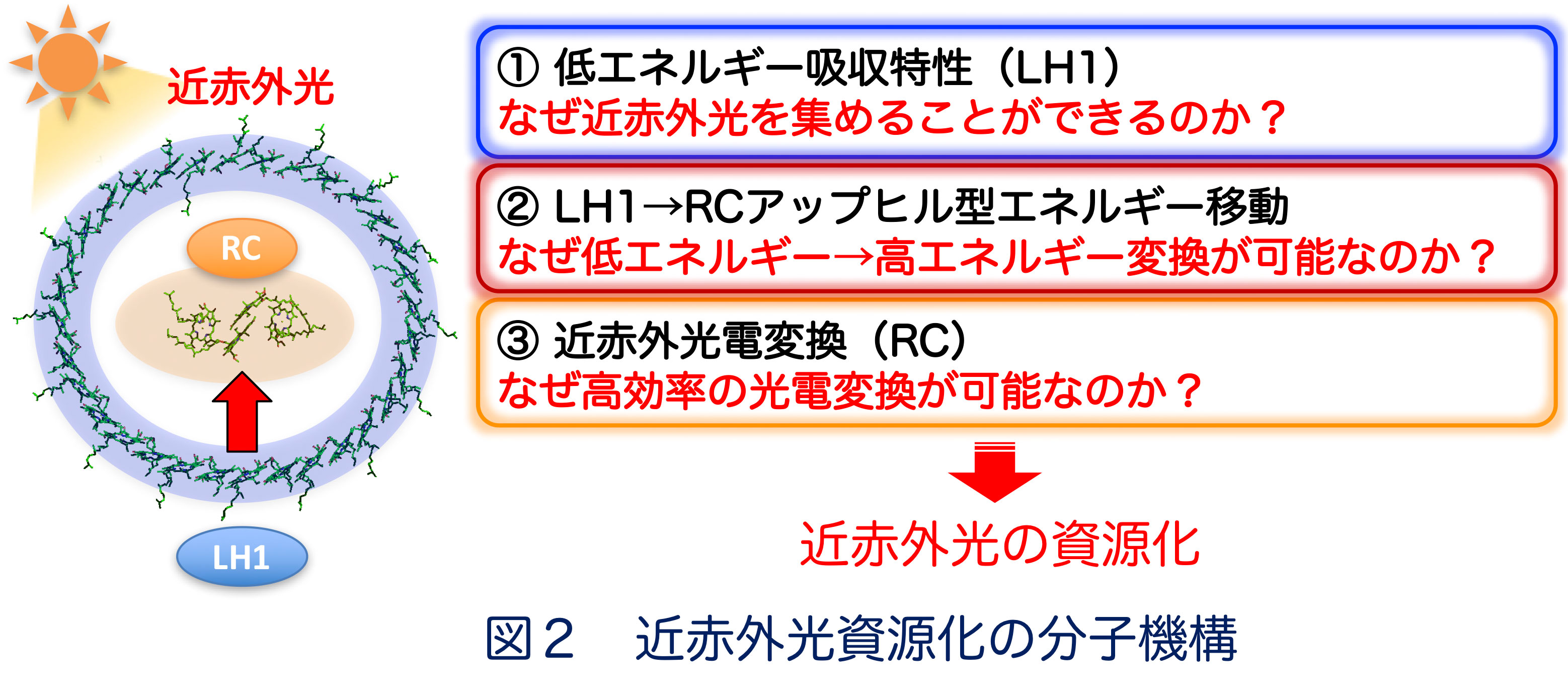
■ 研究シーズ- 光合成細菌による近赤外光のエネルギー資源化シーズカテゴリ:エネルギー, 環境・農学, 自然科学一般研究キーワード:光合成細菌, 近赤外光, 光電変換, アップヒルエネルギー移動研究の背景と目的: 光合成細菌は近赤外光を光電変換する光捕集・電荷分離システム(LH1-RC: 光捕集1反応中心複合体)を備えており、低いエネルギーを高いエネルギーに変換するuphill型エネルギー移動により近赤外光をエネルギー源とした光—物質変換を営んでいます(図1)。本研究では、様々な光捕集特性を示す光合成細菌を用いてuphill型エネルギー移動の分子機構を解明し、近赤外光資源化の基盤創出を目指しています。研究内容: 光合成細菌の光LH1-RCでは、①光捕集アンテナLH1が低エネルギーの近赤外光を吸収し、②LH1から反応中心RCへエネルギー障壁を遡るuphill型エネルギー移動が起こり、③そのエネルギーをRCが電気エネルギーに変換することにより、近赤外光を資源化していると考えられますが、詳細な分子機構は不明です(図2)。 当研究グループでは、様々な環境に棲息する光合成細菌を取り扱っており、光吸収特性やエネルギーギャップが異なるLH1-RC複合体について、それらの構造と機能の関連性を分光学的ならびに構造生物学的な手法により詳しく調べています。 これまでの研究から、①に関して、色素とタンパク質の相互作用、色素分子の種類や配向性、金属イオンの存在などが重要であることが明らかになってきました。現在、②のuphill型エネルギー移動および③の高効率な光電変換のメカニズムを明らかにする研究を進めています。
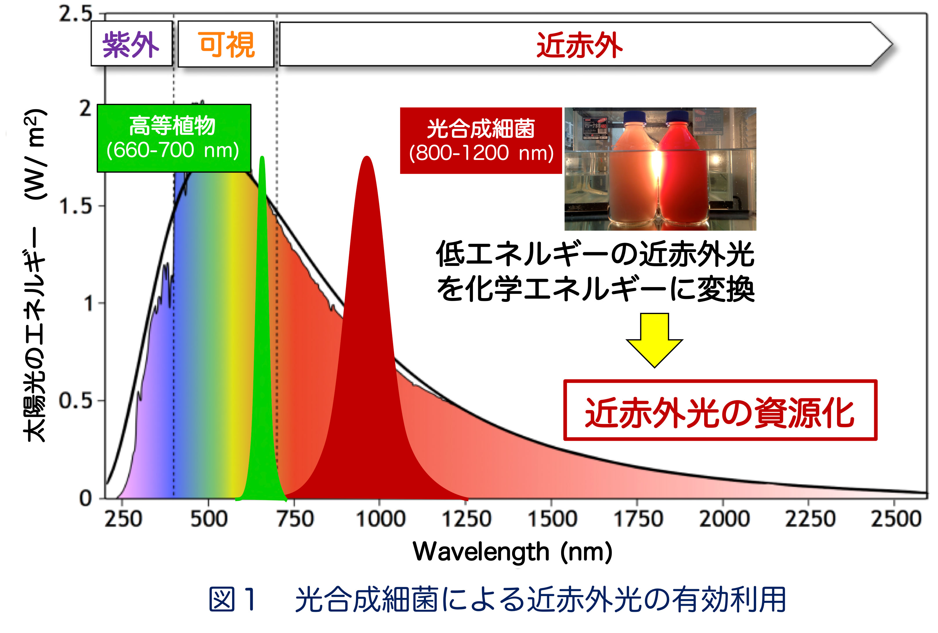
期待される効果や応用分野:図1に示すように、地上に降り注ぐ太陽光の約4割は近赤外光ですが、これらはエネルギーが低いため、未利用のエネルギー資源として浪費されています。光合成細菌はこの近赤外光を利用可能なエネルギーに変換しています。その分子機構の解明は、近赤外光応答型人工光合成系の構築に重要な知見となるものであり、新規エネルギー資源の基盤創出が期待されます。関係する業績:Y. Kimura et al. BBA 1862, 148473, 2021. Y. Kimura et al. Photosynth. Res. 148, 77, 2021. M. Imanishi et al. Biochemistry 58, 2844, 2019. K.V.P. Nagashima et al. PNAS 114, 10906, 2017.